

Encondroma: a propósito de un caso
Palabras clave:
TO- Tumores óseos TOC- Tumores óseos cartilaginosos RT- Resección tumoral TAC- Tomografía axial computarizadaResumen
Fundamento: los tumores óseos son lesiones frecuentes, cada tipo histológico tiene patrones típicos que los identifican. Sin embargo, en ocasiones se pueden presentar de forma atípica.
Objetivo: conocer el caso de una paciente con encondroma en la región proximal de la tibia derecha.
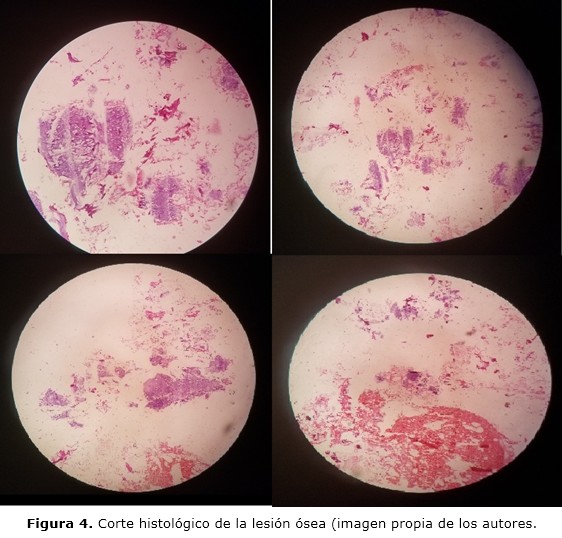
Presentación del caso: mujer de 74 años de edad, blanca con antecedentes de hipertensión arterial y asma bronquial, la cual acude a la consulta de Ortopedia y Traumatología por dolor a nivel de la rodilla derecha que aumenta con la actividad física y se acompaña de limitación funcional y crepitación articular. La tomografía axial computarizada mostró imagen osteoblástica a nivel de la meseta tibial derecha sin ruptura de la cortical. Además se evidenció múltiples quistes subcondrales de aspecto degenerativo, presencia de osteofitos tibiales, femorales ipsi y contralateral, espacio femoro-rotuliano muy disminuido asociado a esclerosis del cóndilo femoral. Con los elementos descritos se decidió llevar la paciente al quirófano, para tratamiento de tipo quirúrgico. Se realizó exéresis de la tumoración, la que fue enviada al departamento de anatomía patológica para estudio histológico que confirmó el diagnóstico. La zona de hueso extraída se llenó con cemento quirúrgico.
Conclusiones: el encondroma es un tumor benigno de origen cartilaginoso, su presencia por encima de los 40 años de edad es ocasional y constituye el principal diagnóstico diferencial del condrosarcoma de bajo grado histológico. Debido a lo infrecuente de esta afección fuera de su rango de edades, es necesaria la biopsia para confirmar el diagnóstico.
DeCS: CONDROMA/diagnóstico; TOMOGRAFÍA COMPUTARIZADA ESPIRAL/métodos; CONDROMA/cirugía; NEOPLASIAS ÓSEAS; INFORMES DE CASOS.
Descargas
Citas
1. Calero-Paniagua I, Vicente-Rodrigo JA, Soliva-Martínez D, Torrecillas-Fernández F. Another Gouty Tophus? The Many Faces of the Enchondroma. Reumatol Clin. 2018;14(4):239-41. DOI: 10.1016/j.reuma.2017.01.002
2. Douis H, Parry M, Vaiyapuri S, Davies AM. What are the differentiating clinical and MRI-features of enchondromas from low-grade chondrosarcomas? Eur Radiol [Internet]. 2018 [citado 08 Nov 2021];28(1):398-409. Disponible en: https://link.springer.com/article/10.1007%2Fs00330-017-4947-0
3. Öztürk R, Arıkan ŞM, Bulut EK, Kekeç AF, Çelebi F, Güngör BŞ. Distribution and evaluation of bone and soft tissue tumors operated in a tertiary care center. Acta Orthop Traumatol Turc [Internet]. 2019 May [citado 08 Nov 2021];53(3):189-94. Disponible en:
https://www.ncbi.nlm.nih.gov/pmc/articles/PMC6599414/.
4. Patel A, Davies AM, Botchu R, James S. A pragmatic approach to the imaging and follow-up of solitary central cartilage tumours of the proximal humerus and knee. Clin Radiol. 2019 Jul; 74(7):517-526. doi: 10.1016/j.crad.2019.01.025
5. Cortés-Cerda R, Mora-Ríos FG, Insunza-Ramírez A, Mejía-Rohenes LC, Ruiz-Alva SK, Pérez García CK. Benign tumors that cause fractures in children. Acta Ortop Mex [Internet]. 2018 Sep-Oct [citado 08 Nov 2021];32(5):283-6. Disponible en: http://www.scielo.org.mx/scielo.php?script=sci_arttext&pid=S2306-41022018000500283&lng=es&nrm=iso&tlng=es
6. Deckers C, Schreuder BHW, Hannink G, de Rooy JWJ, van der Geest IC. Radiologic follow-up of untreated enchondroma and atypical cartilaginous tumors in the long bones. J Surg Oncol. 2016 Dic;114(8):987-91.
7. Holt GE. Evaluation of the patient with bone lesión about the knee. En: Scott WN, editor. Insall & Scott Surgery of the Knee. 6th ed. Philadelphia: Elsevier; 2018. p. 1420.
8. Omlor GW, Lohnherr V, Lange J, Gantz S, Mechtersheimer G, Merle C, et al. Outcome of conservative and surgical treatment of enchondromas and atypical cartilaginous tumors of the long bones: retrospective analysis of 228 patients. BMC Musculoskelet Disord [Internet]. 2019 [citado 08 Nov 2021];20(1):134. Disponible en: https://bmcmusculoskeletdisord.biomedcentral.com/articles/10.1186/s12891-019-2502-7
9. Cable MG, Webber NP, Randall RL. Surgical treatment of benign bone lesions. En: Scott WN editor. Insall & Scott Surgery of the Knee. 6th ed. Philadelphia: Elsevier; 2018.p.2085-95.
10. Afonso PD, Isaac A, Villagrán JM. Chondroid tumors as incidental findings and differential diagnosis between enchondromas and low-grade chondrosarcomas. Semin Musculoskelet Radiol [Internet]. 2019 [citado 08 Nov 2021];23(1):3-18. Disponible en: https://www.thieme-connect.com/products/ejournals/abstract/10.1055/s-0038-1675550
11. Mulligan ME. How to Diagnose enchondroma, bone infarct, and chondrosarcoma. Curr Probl Diagn Radiol. 2019;48(3):262-73. DOI: 10.1067/j.cpradiol.2018.04.002
12. Zhou X, Zhao B, Keshav P, Chen X, Gao W, Yan H. The management and surgical intervention timing of enchondromas: a 10-year experience. Medicine (Baltimore). 2017 Abr;96(16):e6678.
Publicado
Cómo citar
Número
Sección
Licencia
Derechos de autor 2021 Alejandro Alvarez-López, Rodrigo Fuentes-Véjar, Sergio Ricardo Soto-Carrasco, Johenis Creagh-García

Esta obra está bajo una licencia internacional Creative Commons Atribución-NoComercial 4.0.
La Revista Archivo Medico Camagüey, ofrece de forma inmediata después de ser indexada en el Proyecto SciELO; acceso abierto al texto completo de los artículos bajo el principio de hacer disponible y gratuita la investigación para favorecer el intercambio del conocimiento global y coadyuvar a una mayor extensión, publicación, evaluación y uso extensivo de los artículos que se exponen pudiendo ser utilizados, sin fines comerciales, siempre y cuando se haga referencia a la fuente primaria.
Carta De Declaración De Autoría u Derechos De Autor(a)
Conflictos de intereses: los autores deberán declarar de forma obligatoria la presencia o no de conflictos de intereses en relación con la investigación presentada. (Descargar Plantilla para declarar confictos de intereses)
La Revista Archivo Médico Camagüey se encuentra bajo una
Licencia Creative Commons Reconocimiento-NoComercial 4.0 International (CC BY NC 4.0).
Esta licencia permite a otros distribuir, mezclar, ajustar y construir a partir de su obra, incluso con fines comerciales, siempre que le sea reconocida la autoría de la creación original. Esta es la licencia más servicial de las ofrecidas. Recomendada para una máxima difusión y utilización de los materiales sujetos a la licencia. La licencia completa puede consultarse en: https://creativecommons.org/licenses/


