

Encondroma: a propósito de un caso
Keywords:
TO- Tumores óseos TOC- Tumores óseos cartilaginosos RT- Resección tumoral TAC- Tomografía axial computarizadaAbstract
Background: bone tumors are common lesions, there are specific features related to each histological type, but unusual and atypical presentations do occur.
Objective: to show the case of a patient with an enchondroma in the proximal right tibial plateau.
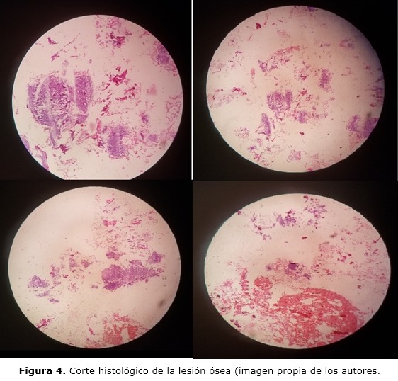
Case report: a 74 year old white woman, who had had previous personal history of hypertension and asthma is taken to the Orthopaedic and Traumatology outpatient department complaining of right knee pain which gets worse with physical activity associated to limited range of motion and articular crepitus. CT scan showed an osteoblastic lesion in the proximal upper right tibial plateau without cortical breaking. On the other hand, multiple subchondral degenerative cysts were found in the knee joint as well as osteophytes, sclerosis and joint space narrowing. Taken into account the entire previous patient' features, surgery was performed and consist of tumor resection with bone cement filled. The specimen was send to the pathology department showing enchondroma.
Conclusions: enchondroma is a benign cartilaginous bone tumor unfrequently found over 40 years of age, the main differential diagnosis is low grade chondrosarcoma. Because of the infrequent presentation of this case in regards to age, surgery was warranted to confirm diagnosis.
DeCS: CHONDROMA/diagnosis; TOMOGRAPHY, SPIRAL COMPUTED/methods; CHONDROMA/surgery; BONE NEOPLASMS; CASE REPORTS.
Downloads
References
1. Calero-Paniagua I, Vicente-Rodrigo JA, Soliva-Martínez D, Torrecillas-Fernández F. Another Gouty Tophus? The Many Faces of the Enchondroma. Reumatol Clin. 2018;14(4):239-41. DOI: 10.1016/j.reuma.2017.01.002
2. Douis H, Parry M, Vaiyapuri S, Davies AM. What are the differentiating clinical and MRI-features of enchondromas from low-grade chondrosarcomas? Eur Radiol [Internet]. 2018 [citado 08 Nov 2021];28(1):398-409. Disponible en: https://link.springer.com/article/10.1007%2Fs00330-017-4947-0
3. Öztürk R, Arıkan ŞM, Bulut EK, Kekeç AF, Çelebi F, Güngör BŞ. Distribution and evaluation of bone and soft tissue tumors operated in a tertiary care center. Acta Orthop Traumatol Turc [Internet]. 2019 May [citado 08 Nov 2021];53(3):189-94. Disponible en:
https://www.ncbi.nlm.nih.gov/pmc/articles/PMC6599414/.
4. Patel A, Davies AM, Botchu R, James S. A pragmatic approach to the imaging and follow-up of solitary central cartilage tumours of the proximal humerus and knee. Clin Radiol. 2019 Jul; 74(7):517-526. doi: 10.1016/j.crad.2019.01.025
5. Cortés-Cerda R, Mora-Ríos FG, Insunza-Ramírez A, Mejía-Rohenes LC, Ruiz-Alva SK, Pérez García CK. Benign tumors that cause fractures in children. Acta Ortop Mex [Internet]. 2018 Sep-Oct [citado 08 Nov 2021];32(5):283-6. Disponible en: http://www.scielo.org.mx/scielo.php?script=sci_arttext&pid=S2306-41022018000500283&lng=es&nrm=iso&tlng=es
6. Deckers C, Schreuder BHW, Hannink G, de Rooy JWJ, van der Geest IC. Radiologic follow-up of untreated enchondroma and atypical cartilaginous tumors in the long bones. J Surg Oncol. 2016 Dic;114(8):987-91.
7. Holt GE. Evaluation of the patient with bone lesión about the knee. En: Scott WN, editor. Insall & Scott Surgery of the Knee. 6th ed. Philadelphia: Elsevier; 2018. p. 1420.
8. Omlor GW, Lohnherr V, Lange J, Gantz S, Mechtersheimer G, Merle C, et al. Outcome of conservative and surgical treatment of enchondromas and atypical cartilaginous tumors of the long bones: retrospective analysis of 228 patients. BMC Musculoskelet Disord [Internet]. 2019 [citado 08 Nov 2021];20(1):134. Disponible en: https://bmcmusculoskeletdisord.biomedcentral.com/articles/10.1186/s12891-019-2502-7
9. Cable MG, Webber NP, Randall RL. Surgical treatment of benign bone lesions. En: Scott WN editor. Insall & Scott Surgery of the Knee. 6th ed. Philadelphia: Elsevier; 2018.p.2085-95.
10. Afonso PD, Isaac A, Villagrán JM. Chondroid tumors as incidental findings and differential diagnosis between enchondromas and low-grade chondrosarcomas. Semin Musculoskelet Radiol [Internet]. 2019 [citado 08 Nov 2021];23(1):3-18. Disponible en: https://www.thieme-connect.com/products/ejournals/abstract/10.1055/s-0038-1675550
11. Mulligan ME. How to Diagnose enchondroma, bone infarct, and chondrosarcoma. Curr Probl Diagn Radiol. 2019;48(3):262-73. DOI: 10.1067/j.cpradiol.2018.04.002
12. Zhou X, Zhao B, Keshav P, Chen X, Gao W, Yan H. The management and surgical intervention timing of enchondromas: a 10-year experience. Medicine (Baltimore). 2017 Abr;96(16):e6678.
Published
How to Cite
Issue
Section
License
Copyright (c) 2021 Alejandro Alvarez-López, Rodrigo Fuentes-Véjar, Sergio Ricardo Soto-Carrasco, Johenis Creagh-García

This work is licensed under a Creative Commons Attribution-NonCommercial 4.0 International License.
Copyright: Camagüey Medical Archive Magazine, offers immediately after being indexed in the SciELO Project; Open access to the full text of the articles under the principle of making available and free the research to promote the exchange of global knowledge and contribute to a greater extension, publication, evaluation and extensive use of the articles that can be used without purpose As long as reference is made to the primary source.
Conflicts of interest: authors must declare in a mandatory manner the presence or not of conflicts of interest in relation to the investigation presented.
(Download Statement of potential conflicts of interest)
The Revista Archivo Médico de Camagüey is under a License Creative Commons Attribution-Noncommercial-No Derivative Works 4.0 International (CC BY 4.0).
This license allows others to distribute, to mix, to adjust and to build from its work, even for commercial purposes, as long as it is recognized the authorship of the original creation. This is the most helpful license offered. Recommended for maximum dissemination and use of licensed materials. The full license can be found at: https://creativecommons.org/licenses/


 22 julio 2025