Síndrome doloroso de la fabela
Resumen
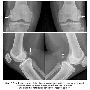
Introducción: La fabela es un hueso sesamoideo inconstante localizado en la cara posterolateral de la rodilla, en ocasiones está involucrado en la presencia de dolor y trastornos acroparestésicos.
Objetivo: Actualizar y brindar información sobre los aspectos más importantes del síndrome doloroso de la fabela.
Métodos: La búsqueda y análisis de la información se realizó en un periodo de 91 días (primero de septiembre al 30 de noviembre de 2024) y se emplearon las siguientes palabras: Fabella pain syndrome, fabella AND posterolateral knee pain, fabella, fabellectomy, arthroscopy. A partir de la información obtenida se realizó una revisión bibliográfica de un total de 171 artículos publicados en las bases de datos PubMed, Hinari, SciELO, ResearchGate, Ebsco, Scopus, Medscape y Medline mediante el gestor de búsqueda y administrador de referencias EndNote, de ellos se utilizaron 31 citas seleccionadas para realizar la revisión, 30 de los últimos cinco años.
Resultados: Se hace referencia a la anatomía de la zona, las diferencias entre fabela y ciamela y de los estabilizadores estáticos y dinámicos. Se abordan los aspectos clínicos, imagenológicos y electromiográficos. Con relación al tratamiento se describen las modalidades conservadoras y quirúrgicas con sus principales indicaciones, con especial énfasis en la cirugía artroscópica.
Conclusiones: El síndrome doloroso de la fabela se caracteriza por dolor en la cara posterolateral de la rodilla que aumenta con la actividad física en especial la extensión. Los métodos imagenológicos son esenciales para el diagnóstico de esta enfermedad. Las modalidades de tratamiento son conservadora o quirúrgica, este último consiste en la resección de la fabela con o sin descompresión del nervio peroneo común.
DeCS: RODILLA/diagnóstico; ARTROSCOPÍA; DOLOR MUSCULOESQUELÉTICO; FACTORES DE RIESGO; REVISIÓN.
Descargas
Citas
1. Charalbous CP. The Knee Made Easy [Internet]. Nueva York: Springer; 2022 [citado 30 Mar 2024]. Disponible en: https://doi.org/10.1007/978-3-030-54506-2
2. Sodavarapu P, Kumar D, Guduru A, Rathod PM. An Unusual Complex Posterolateral Osteoligamentous Injury of the Knee in an Adolescent. Cureus [Internet]. 2020 [citado 30 Mar 2024];12(4): e7532. Disponible en: https://doi.org/10.7759/cureus.7532
3. Xu L, Wei YK, Jiao HB, Song YC. Relationship between fabella and posterolateral knee pain and common peroneal nerve injury. Zhongguo Gu Shang [Internet]. 2020 [citado 30 Mar 2024];33(11):1071-5. Disponible en:
https://doi.org/10.12200/j.issn.1003-0034.2020.11.017
4. Diaz CC, Agarwalla A, Forsythe B. Fabella syndrome and common peroneal neuropathy following total knee arthroplasty. Case Rep Orthop [Internet]. 2021 [citado 30 Mar 2024]:7621844. Disponible en: https://doi.org/10.1155/2021/7621844
5. Kimura T, Tanikawa H, Hasegawa T, Takeda K, Harato K, Kobayashi S; et al. Late onset of the fabella syndrome after total knee arthroplasty. Case Rep Orthop [Internet]. 2019 [citado 30 Mar 2024]: 5219237. Disponible en:: https://doi.org/10.1155/2019/5219237
6. Provencher MT, Sanchez G, Ferrari MB, Moatshe G, Chahla J, Akamefula R; et al. Artrhoscopy assited fabella excision: surgical technique. Arthros Tech [Internet]. 2017 [citado 30 Mar 2024]; 6(2): e369-e374. Disponible en: http://doi.org/10.1016/j.eats.2016.10.011
7. Ernat JJ, Peebles AM, Provencher MT. Open excision of a painful fabella. Arthrosc Tech [Internet]. 2022 [citado 30 Mar 2024];11(4): e577-e581. Disponible en: https://doi.org/10.1016/j.eats.2021.12.010
8. Asghar A, Naaz S, Chaudhary B. The ethnic and geographical distribution of fabella: a systematic review and meta-analysis of 34,733 knees. Cureus [Internet]. 2021 [citado 30 Mar 2024];13(4):e14743. Disponible en: https://doi.org/10.7759/cureus.14743
9. Zhong J, Zhang G, Si L, Hu Y, Xing Y, He Y; et al. The prevalence and parameters of fabella and its association with medial meniscal tear in China: a retrospective study of 1011 knees. BMC Musculoskelet Disord [Internet]. 2022 [citado 30 Mar 2024];23(1):188. Disponible en: https://doi.org/10.1186/s12891-022-05092-4
10. Akdeniz H, Ozkan S, Adanas C. Prevelance of fabella: an MRI study in The Eastern Anatolia Region of Turkey. Curr Med Imaging [Internet]. 2021 [citado 30 Mar 2024];17(10):1221-1225. Disponible en: https://doi.org/10.2174/1573405617666210528121352
11. Dekker TJ, Crawford MD, DePhillipo NN, Kennedy MI, Grantham WJ, Schairer WW; et al. Clinical presentation and outcomes associated with fabellectomy in the setting of fabella syndrome. Orthop J Sports Med [Internet]. 2020 [citado 30 Mar 2024];8(2):2325967120903722. Disponible en: https://doi.org/10.1177/2325967120903722
12. Akkoc RF, Aksu F, Emre E, Sap O, Karatas A, Aydin S; et al. The morphology of fabella and its prevalence in Turkish society. Eur Rev Med Pharmacol Sci [Internet]. 2022 [citado 30 Mar 2024];26(4):1164-1169. Disponible en: https://doi.org/10.26355/eurrev_202202_28108
13. Berthaume MA, Bull AMJ. Human biological variation in sesamoid bone prevalence: the curious case of the fabella. J Anat [Internet]. 2020 [citado 30 Mar 2024];236(2):228-242. Disponible en: https://doi.org/10.1111/joa.13091
14. Aksu F, Akkoc RF, Gundogan Bozdag P. The morphology of cyamella and its prevalence in Turkish society. Eur Rev Med Pharmacol Sci [Internet]. 2023 [citado 30 Mar 2024];27(19):9085-9090. Disponible en: https://doi.org/10.26355/eurrev_202310_33934
15. Basaran S, Coskun Benlidayi I. Coexistence of symptomatic cyamella and multiple fabellae: a case report. Arch Rheumatol [Internet]. 2022 [citado 30 Mar 2024];38(1):156-158. Disponible en: https://doi.org/10.46497/ArchRheumatol.2022.9521
16. Cheppalli NS, Purudappa PP, Price R, Kolwadkar Y, Metikala S. Isolated lateral-sided knee pain in total knee arthroplasty. A review. Orthop Rev (Pavia) [Internet]. 2024 [citado 30 Mar 2024]; 16:93014. Disponible en: https://doi.org/10.52965/001c.93014
17. Özbay H, Mraja HM, Can A, Erdogan F. Prevalence and radiological characteristics of the fabella in Turkish population. Cureus [Internet]. 2022 [citado 30 Mar 2024];14(11):e31534. Disponible en: https://doi.org/10.7759/cureus.31534
18. Hou W, Xu L, Wang J, Wang B, Liu L, Xu K; et al. Fabellar prevalence, degeneration and association with knee osteoarthritis in the Chinese population. Sci Rep [Internet]. 2019 [citado 30 Mar 2024];9(1):13046. Disponible en: https://doi.org/10.1038/s41598-019-49174-1
19. Lin JC, Tsai MH, Lin WP, Kuan TS, Lien WC. Entrapment neuropathy of common peroneal nerve by fabella: a case report. World J Clin Cases [Internet]. 2023 [citado 30 Mar 2024];11(28):6857-6863. Disponible en: https://doi.org/10.12998/wjcc.v11.i28.685
20. Loscos S, López-Vidriero R, López-Vidriero E. Fabella syndrome in an elite swimmer. Rev Esp Cir Ortop Traumatol (Engl Ed) [Internet]. 2020 [citado 30 Mar 2024];64(5):361-364. Disponible en: https://doi.org/10.1016/j.recot.2020.04.008
21. Wolinski F, Brylinski L, Kostelecka K, Teresinski G, Buszewicz G, Baj J. Common fibular nerve palsy due to the fabella: a review. Clin Anat [Internet]. 2024 [citado 30 Mar 2024];37(1):73-80. Disponible en: https://doi.org/10.1002/ca.24089
22. Buruian A, Pinheiro V, Fonseca F, Matos P. Fracture of the fabella with radiologic and MRI. BMJ Case Rep [Internet]. 2023 [citado 30 Mar 2024];16(11):e251811. Disponible en: https://doi.org/10.1136/bcr-2022-251811
23. Matroushi ODA, Sirasanagandla SR, Shabibi AA, Obaidani AA, Dhuhli HA, Jaju S; et al. Radiological study of fabella in Omani subjects at a tertiary care center. Anat Cell Biol [Internet]. 2021 [citado 30 Mar 2024];54(3):315-320. Disponible en: https://doi.org/10.5115/acb.20.194
24. Adedigba JA, Idowu BM, Hermans SP, Okwori OF, Onigbinde SO, Oluwadiya KS; et al. Fabella and patella variants: radiographic prevalence, distribution and clinical relevance in a population of black african descent. Anat Cell Biol [Internet]. 2021 [citado 30 Mar 2024];54(2):184-192. Disponible en: https://doi.org/10.5115/acb.20.217
25. Li YM, Kao CL. Sonographic diagnosis and treatment of fabella syndrome: a neglected posterolateral knee pain. Am J Phys Med Rehabil [Internet]. 2023 [citado 30 Mar 2024];102(2): e23-e24. Disponible en: https://doi.org/10.1097/PHM.0000000000002091
26. Pekala PA, Mann MR, Pekala JR, Loukas M, Wojciechowski W, Walocha JA; et al. The gastrocnemiofibular ligament: a new, more anatomically accurate name for the fabellofibular ligament-an original magnetic resonance imaging study and meta-analysis. Clin Anat [Internet]. 2020 [citado 30 Mar 2024];33(3):419-427. Disponible en: https://doi.org/10.1002/ca.23542
27. Unluturk O, Duran S, Yasar Teke H. Prevalence of the fabella and its general characteristics in Turkish population with magnetic resonance imaging. Surg Radiol Anat [Internet]. 2021 [citado 30 Mar 2024];43(12):2047-2054. Disponible en: https://doi.org/10.1007/s00276-021-02817-3
28. Hur JW, Lee S, Jun JB. The prevalence of fabella and its association with the osteoarthritic severity of the knee in Korea. Clin Rheumatol [Internet]. 2020 [citado 30 Mar 2024];39(12):3625-3629. Disponible en: https://doi.org/10.1007/s10067-020-05078-4
29. Nguyen DQ, Do TD, Van Nguyen L, Mai VD, Do CD. Fabella syndrome in a professional football player: a case report and literature review. Int J Surg Case Rep [Internet]. 2022 [citado 30 Mar 2024]; 93:106919. Disponible en: https://doi.org/10.1016/j.ijscr.2022.106919
30. Samra D, Cross T, Feller J, Gultekin S. Outcome of fabellar excision on return to sport and performance for an elite athlete with established lateral compartment chondropathy. Orthop J Sports Med [Internet]. 2021 [citado 30 Mar 2024];9(9):23259671211034157. Disponible en:: https://doi.org/10.1177/23259671211034157
31. Weng SP, Wu TM, Chien CS, Lin SH. Treatment of fabella syndrome with arthroscopic fabellectomy: a case series and literature review. BMC Musculoskelet Disord [Internet]. 2021 [citado 30 Mar 2024];22(1):748. Disponible en: https://doi.org/10.1186/s12891-021-04630-w
Publicado
Cómo citar
Número
Sección
Licencia
La Revista Archivo Medico Camagüey, ofrece de forma inmediata después de ser indexada en el Proyecto SciELO; acceso abierto al texto completo de los artículos bajo el principio de hacer disponible y gratuita la investigación para favorecer el intercambio del conocimiento global y coadyuvar a una mayor extensión, publicación, evaluación y uso extensivo de los artículos que se exponen pudiendo ser utilizados, sin fines comerciales, siempre y cuando se haga referencia a la fuente primaria.
Carta De Declaración De Autoría u Derechos De Autor(a)
Conflictos de intereses: los autores deberán declarar de forma obligatoria la presencia o no de conflictos de intereses en relación con la investigación presentada. (Descargar Plantilla para declarar confictos de intereses)
La Revista Archivo Médico Camagüey se encuentra bajo una
Licencia Creative Commons Reconocimiento-NoComercial 4.0 International (CC BY NC 4.0).
Esta licencia permite a otros distribuir, mezclar, ajustar y construir a partir de su obra, incluso con fines comerciales, siempre que le sea reconocida la autoría de la creación original. Esta es la licencia más servicial de las ofrecidas. Recomendada para una máxima difusión y utilización de los materiales sujetos a la licencia. La licencia completa puede consultarse en: https://creativecommons.org/licenses/