

Asociación entre apiñamiento anteroinferior y tercer molar en pacientes de 20 años de edad
Resumen
Introducción: El apiñamiento dentario anteroinferior es una maloclusión frecuente en el ser humano. Entre sus causas se cita al tercer molar inferior, sin embargo, no existe suficientes estudios que avalen una dependencia estadística entre el apiñamiento y el tercer molar.
Objetivo: Determinar la asociación entre el apiñamiento anteroinferior y el tercer molar inferior.
Métodos: Se realizó un estudio descriptivo transversal, en el que participaron 68 estudiantes de 20 años de la facultad de Estomatología de la provincia Camagüey, durante el período de noviembre de 2019 a febrero de 2020. Se determinó presencia del tercer molar y de apiñamiento; así como, posición del tercer molar y espacio para su ubicación en el arco.
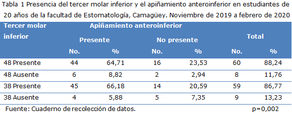
Resultados: Hubo supremacía en la existencia del tercer molar inferior en pacientes estudiados, donde se presentó en su mayoría el apiñamiento anteroinferior. La posición no adecuada predominó en ambos molares inferiores, lo que coincide con una mayor prevalencia de apiñamiento. El espacio para el molar 48 fue en su generalidad suficiente, mientras para el diente 38 fue el no existe; el apiñamiento se diagnosticó tanto donde hubo el espacio suficiente como en los que estuvo o no existió. Se encontró asociación estadística entre el apiñamiento anteroinferior y el tercer molar inferior con posición no adecuada.
Conclusiones: Se constató que los terceros molares inferiores estuvieron presentes en la mayoría de los casos estudiados lo que coincide con la mayor parte de los pacientes con apiñamiento anteroinferior.
DeCS: TERCER MOLAR; MALOCLUSIÓN; ERUPCIÓN DENTAL; DIENTE; ODONTOLOGÍA.
Descargas
Citas
REFERENCIAS BIBLIOGRÁFICAS
1. García Carpio GA.Prevalencia de apiñamiento dental asociado a terceros molares en clínicas de Facultad Piloto De Odontología [tesis].Guayaquil: Universidad de Guayaquil; 2020[citado 28Mar 2022]. Disponible en: http://repositorio.ug.edu.ec/bitstream/redug/48542/1/3229GARCIAgustavo.pdf
2. Kiep P, Duerksen G, Cantero L, López A, Núñez Mendieta H, Ortiz R, Keim L. Grado de maloclusiones según el índice de estética dental en pacientes que acudieron a la Universidad del Pacífico. Rev. cient. cienc. salud[Internet]. 2021 [citado 28Mar 2022]; 3(1):[aprox.7p.].Disponible en:
Doi: 10.53732/rccsalud/03.01.2021.56
3. González Amaral MG, Rodríguez López LV. Prevalencia, tipos y factores etiológicos de apiñamiento mandibular tardío en pacientes de ortodoncia en Tabasco, México, 2015-2016. RevMex de Ortodoncia [Internet]. 2018 [citado 2022 Mar 28]; 6(1):[aprox.7p.]. Disponible en:
http://www.medigraphic.com/ortodoncia
4. Suárez-González M de la C, Gutiérrez-Olives Y, Navarro-Nápoles J, Núñez Oduardo EA, Rosales-Ramírez Y. Maloclusiones dentarias en escolares según índice de estética dental. Rev Electrónica Dr. Zoilo E. MarinelloVidaurreta [Internet]. 2020 Sep-Oct [citado 28 Mar 2022]; 45(5):[aprox.5p.]. Disponible en: http://revzoilomarinello.sld.cu/index.php/zmv/article/view/2203
5. Bustillo Arrieta J. Implicación de la erupción de los terceros molares en el apiñamiento anteroinferior severo. AvOdontoestomatol [Internet]. 2016 Mar-Abr [citado 28 Mar 2022]; 32(2): [aprox.5p.]. Disponible en: http://scielo.isciii.es/scielo.php?script=sci_arttext&pid=S0213-12852016000200005&lng=es.
6. Cuellar Gutiérrez J, Begoña Moreno, Muñoz M, Veloso Bustos D, Villanueva J. Relación entre apiñamiento dentario y terceros molares. Rev. Clin. Periodoncia Implantol. Rehabil. Oral [Internet]. 2018 Dic [citado 16 Feb 2022]; 11(3):[aprox.3p.]. Disponible en: http://dx.doi.org/10.4067/S0719-01072018000300173
7. Palacios Vivar DE, LlanesSerantes M, Calderón Lumbreras A, Pérez Aguilar EY, Paredes Tenesaca DG. Predicción del tercer molar e indicaciones de germenectomía. Reporte de un caso. Revista ADM [Internet]. 2018 Dic [citado 14 Feb 2022]; 75(5): [aprox.6p.]. Disponible en: http://eds.b.ebscohost.com/abstract?site=eds&scope=site&jrnl=00010944&AN=134832042&h=drIYQP4Sj9E9VkAsvuGzIlxz%2f%2ftQ1A5%2b77Wklejs6LYrhdOfiAkRG7roFySbiswZKr6vxXLvXdOOi9%2b8KNdc1w%3d%3d&crl=c&resultLocal=ErrCrlNoResults&resultNs=Ehost&crlhashurl=login.aspx%3fdirect%3dtrue%26profile%3dehost%26scope%3dsite%26authtype%3dcrawler%26jrnl%3d00010944%26AN%3d134832042
8. Gutiérrez L. Apiñamiento dental: sus causas, consecuencias y mejores tratamientos. Dentaly.org [actualizado 23 May 2019; citado10 Feb 2022]. [aprox 2 pantallas].Disponible en: https://www.dentaly.org/es/ortodoncia/maloclusion/apinamiento-dental/
9. NavarroAsencioE, JiménezGarcíaE, RappoportRedondoS&Thoilliez Ruano B. Fundamentos de la investigación y la innovación educativa. La Rioja, Spain: Unir Editorial, 2017.
10. Fuentes FR, Borie EE, BustosML, Thomas MD. Morfometría de terceros molares: un estudio de 55 casos. Int. J. Morphol[Internet]. 2009 Dic [citado 14 Feb 2022];27(4): [aprox.6p.]. Disponible en:
https://dx.doi.org/10.406/s0717-95022009000400050
11. Carbo Ayala JE. Anatomía dental y de la oclusión. La Habana: Editorial Ciencias Médicas; 2009.
12. Olguín Martínez T, Amarillas Escobar ED. Morfología Radicular de los Terceros Molares. ADM[Internet].2017[citado 11 Feb 2022]; 74(7): [aprox.6p.]. Disponible en: https://pesquisa.bvsalud.org/portal/resource/pt/biblio-869348
13. González EL. Características anatomorradiográficas de los terceros molares en adolescentes de la enseñanza preuniversitaria. Rev Cubana Estomatol[Internet].2019[citado 11 Feb 2022]; 56(2):[aprox.13p.]. Disponible en:
https://wwwmedigraphic.com/cgi.bin/new-resumen.cgi?IDARTICULO=90837
14. Richardson ME. Lower third molar development subsequent to second molar extraction. Am. J. Orthod. Dentofac. Orthop[Internet]. 1993[citado 11 Feb 2022]; 104(6):[aprox. 9 p.].Disponible en:https://www.sciencedirect.com/science/article/abs/pii/S0889540605804408
15. Jiménez Sánchez AC, Sierra- Robles E. Frecuencia de agenesias dentales en pacientes q acudieron a un centro radiológico en Guadalajara, México. RevTamé"[Internet].2019[citado 11 Feb 2022]; 8(22): [aprox.6p.]. Disponible en:
http://www.medigraphic.com/cgi-bin/new/publicaciones.cgi?IDREVISTA=342
16. González Amaral MG, Rodríguez López LV. Prevalencia, tipos y factores etiológicos de apiñamiento mandibular tardío en pacientes de ortodoncia en Tabasco, México, 2015-2016. Rev. Mex. Ortodoncia [Internet]. 2018 Ene [citado 16 Feb 2022]; 6(1): [aprox.6p.]. Disponible en:http://www.medigraphic.org.mx
17. González Espangler L, rodríguez Torres E, Soto Cantero LA, Romero García LI, Pichel Borges I. Modificaciones del espacio óseo posterior para terceros molares desde la infancia hasta la adolescencia. MEDISAN[Internet]. 2019 Sep-Oct [citado 15 Feb 2022]; 23(5): [aprox.5p.]. Disponible en: http://www.redalyc.org/articulo.oa?id=368461459007
18. Mosquera-Valencia Y, Vélez-Zapata D, Velásquez-Velásquez M. Frecuencia de posiciones de terceros molares impactados en pacientes atendidos en la IPS. Rev. CES Odont [Internet].2020[citado 11 Feb 2022];33(1):[aprox.7p.].Disponible en: http://www.scielo.org.co/scielo.php?script=sci_arttext&pid=S0120-971X2020000100022
19. Gökçe G, Akan B, Veli I. El papel de la angulación del tercer molar impactado en el apiñamiento anterior. Ortodoncia de tendencias APOS [Internet]. 2021 [citado 15 Feb 2022]; 11(1):[aprox.4p.]. Disponible en: http://www.researchgate.net
20. Restrepo Rendón LF, Meneses Tamayo F, Vivares Builes AM. Complicaciones quirúrgicas y posquirúrgicas en la exodoncia de terceros molares inferiores: estudioretrospectivo. Acta Odontológica Colombiana [Internet]. 2019 Ene [citado 11 Feb 2022]; 9(1):[aprox.11p.]. Disponible en:https://revistas.unal.edu.co/index.php/actaodontocol/article/view/72842
21. Acosta Rodríguez A, Morales Navarro D, Cárdenas Moya J.Grado de dificultad en terceros molares mandibulares retenidos. Archivos del Hospital Universitario "General Calixto García"[Internet].2021[citado 11 Feb 2022]; 9(1):[aprox.6p.]. Disponible en:
http://www.revcalixto.sld.cu/index.php/ahcg/article/view/e614/596
22. Mayoral G, Mayoral J. Ortodoncia: Principios fundamentales y prácticos.Ciudad de La Habana:Editorial Cientifico-Técnica;1986.
23. Pino Román IM, Álvarez Martínez OL, Benavides Sosa Y, Fuentes González Y, García Rodríguez M. Malocclusions according to the Dental Aesthetic Index (DAI) in 7th grade students. Acta méd [Internet]. 2020[citado 15 Feb 2022]; 14(3):[aprox.4p.]. Disponible en: http://scielo.sld.cu/scielo.php?script=sci_arttex&pid=s2709-79272020000300357
24. Gutiérrez-Rojo JF, Gutiérrez-Villaseñor J, Mú-Gálvez BY, Navarrete Ayón KB, García Rivera RN. Frecuencia de dientes con erupción ectópica de la Especialidad de Ortodoncia de la Universidad Autónoma de Nayarit. RevTame [Internet]. 2019[citado15 Feb 2022];8(23): [aprox.3p.]. Disponible en:
https://www.medigraphic.com/cgi-bin/new/resumenI.cgi?IDARTICULO=91289
25. Rosero López JC. Factores de riesgo en exodoncia del tercer molar inferior[tesis].Ecuador: Universidad de Guayaquil. Facultad Piloto de Odontología; 2020 [citado15 Feb 2022].Disponible en: http://repositorio.ug.edu.ec/handle/redug/48531
Publicado
Cómo citar
Número
Sección
Licencia
Derechos de autor 2022 Nereisy Mirabal-García, Eliane Leyva-Arango, Norys Tan-Suárez, Thalía Machado-Tan, Idalmis Reytor-González, Estrella de la Noval-Gómez

Esta obra está bajo una licencia internacional Creative Commons Atribución-NoComercial 4.0.
La Revista Archivo Medico Camagüey, ofrece de forma inmediata después de ser indexada en el Proyecto SciELO; acceso abierto al texto completo de los artículos bajo el principio de hacer disponible y gratuita la investigación para favorecer el intercambio del conocimiento global y coadyuvar a una mayor extensión, publicación, evaluación y uso extensivo de los artículos que se exponen pudiendo ser utilizados, sin fines comerciales, siempre y cuando se haga referencia a la fuente primaria.
Carta De Declaración De Autoría u Derechos De Autor(a)
Conflictos de intereses: los autores deberán declarar de forma obligatoria la presencia o no de conflictos de intereses en relación con la investigación presentada. (Descargar Plantilla para declarar confictos de intereses)
La Revista Archivo Médico Camagüey se encuentra bajo una
Licencia Creative Commons Reconocimiento-NoComercial 4.0 International (CC BY NC 4.0).
Esta licencia permite a otros distribuir, mezclar, ajustar y construir a partir de su obra, incluso con fines comerciales, siempre que le sea reconocida la autoría de la creación original. Esta es la licencia más servicial de las ofrecidas. Recomendada para una máxima difusión y utilización de los materiales sujetos a la licencia. La licencia completa puede consultarse en: https://creativecommons.org/licenses/


