

Cranio-spinal length: predictor of fetal growth and the trophic condition of the newborn
Abstract
Background: age and fetal growth are determined by head length or cranial-spinal length between weeks 5 and 10 of gestation and then a combination of measurements of other biometric variables is used.
Objective: to identify the correlation of biometric variables and trophic condition at birth with the cranial-spinal length of the first trimester.
Methods: an investigation with a retrospective longitudinal analytical design was carried out in Villa Clara province; it was appealed in the period from January 2013 to October 2018. The population consisted of 6050 pregnant women. The sample was made through an intentional non-probabilistic sampling by criteria, consisting of 3 910 pregnant women. Data were obtained from genetics consultation record books from selected health areas. In the analysis, Pearson's linear correlation coefficient, Spearman's Rho, and scatter diagrams were used.
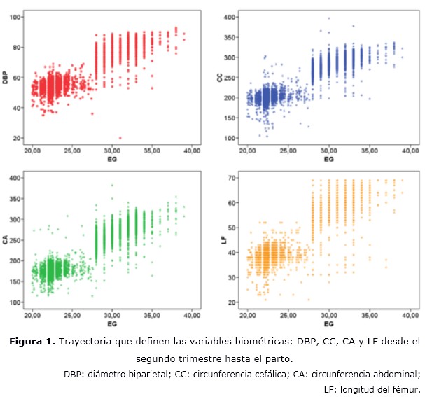
Results: there was a correlation between the cranial-spinal length with the variables and with the trophic condition of the newborn. The graphs that defined the trajectory of the biometric variables from the second trimester to delivery showed compaction of the values up to week 25 and dispersion between week 25 and 30 with differential features in them.
Conclusions: the correlation found of cranial-spinal length with biometric variables and with the trophic condition of the newborn demonstrates the usefulness of the use of this variable in the evolution of pregnancy by both the obstetrician and the comprehensive general practitioner; particularly in pregnant women in whom some growth alteration is suspected. The results obtained motivate further study by subdividing the sample into subgroups such as those affected by diabetes mellitus or arterial hypertension or another, particularly because there is no information on it in the literature.
DeCS: PREGNANCY; BIOMETRY; ULTRASONOGRAPHY; EMBRYONIC AND FETAL DEVELOPMENT; INFANT, NEWBORN/growth&development.
Downloads
References
1. Puig Palau A. Evaluación del crecimiento posnatal en los prematuros de muy bajo peso con edad gestacional menor o igual a 32 semanas desde el nacimiento hasta los 5 años de vida [Tesis Doctoral]. España: Universidad Autónoma de Barcelona; 2017 [citado 05 Jun 2020]. Disponible en: https://www.tdx.cat/bitstream/handle/10803/457736/app1de1.pdf?sequence=1&isAllowed=y
2. Paladino Castillo NN. Diagnóstico y Seguimiento del feto con Restricción del Crecimiento Intrauterino mediante la Aplicación Clínica de la Flujometría Doppler en gestantes del Hospital Bertha Calderón Roque de Abril 2014–Enero 2017 [Tesis]. Managua: Universidad Nacional Autónoma de Nicaragua; 2017 [citado 05 Jun 2020]. Disponible en: https://repositorio.unan.edu.ni/4531/1/96680.pdf
3. Kliegman RM, Geme JS, Blum N, Shah SS, Tasker RC. 21 ed. Nelson. Tratado de Pediatría. España: Elsevier Health Sciences; 2020.
4. Barker DJP. Past obstacles and future promise [Internet]. 2006 [citado 05 Jun 2020]. Disponible en: https://www.researchgate.net/publication/312537369_Past_obstacles_and_future_promise
5. Alban Bautista EP, Zapata Cornejo PG. Coeficiente de concordancia en la estimación del ponderado fetal en gestantes mediante ecografía comparado con regla de Johnson Tumbes 2017 [Tesis]. Tumbes: Universidad Nacional de Tumbes; 2017 [citado 05 Jun 2020]. Disponible en: http://repositorio.untumbes.edu.pe/bitstream/handle/20.500.12874/81/TESIS%20-%20ALBAN%20Y%20ZAPATA.pdf?sequence=1&isAllowed=y
6. Limas Pérez Y, Álvarez-Guerra González E, Sarasa Muñoz N, Cañizares Luna O, Artiles Santana A, Machado Díaz B. Efectividad de los indicadores antropométricos para el diagnóstico de restricción del crecimiento intrauterino. Rev cuba obstet ginecol [Internet]. 2019 [citado 05 Mar 2020];45(1). Disponible en: http://revginecobstetricia.sld.cu/index.php/gin/article/view/418
7. Mejía Salazar AM. Eficacia de la estimación de peso fetal por mediciones ecográficas sobre la macrosomía fetal [Tesis Maestría]. Guatemala: Universidad de San Carlos; Jun 2016 [citado 05 Jun 2020]. Disponible en: http://biblioteca.usac.edu.gt/tesis/05/05_10154.pdf
8. Sadller TW. Langman. Embriología médica. 14 ed. Paris: Lippincott Williams & Wilkins; 2019.
9. Subcomisiones, Comités y Grupos de Trabajo. Propuesta de actualización de la evaluación antropométrica del recién nacido. Arch Argent Pediatr [Internet]. 2017 [citado 05 Jun 2020];115(1):[aprox. 7 p.]. Disponible en: https://www.researchgate.net/publication/314039702_Propuesta_de_actualizacion_de_la_evaluacion_antropometrica_del_recien_nacido_Proposal_to_update_the_anthropometric_evaluation_of_the_newborn
10. Menéndez Pedraja Y, Mojena Roblejo M, Estrada López K, Bravet Smith E, Mojena Medina D. Valores biométricos fetales y peso fetal estimado en el tercer trimestre de la gestación [Internet]. Manzanillo, Granma: Primer Congreso Virtual de Ciencias Básicas Biomédicas en Granma; 2020 [citado 23 Nov 2020]. Disponible en: http://cibamanz2020.sld.cu/index.php/cibamanz/cibamanz2020/paper/viewFile/419/223
11. Pérez Julca LG. Método ecográfico versus método clínico en la predicción del peso fetal de gestantes a término del servicio de obstetricia del hospital nacional Alberto Sabogal Sologuren-2018 [Tesis]. Lima: Universidad Nacional Federico Villarreal; 2019 [citado 05 Jun 2020]. Disponible en: http://repositorio.unfv.edu.pe/bitstream/handle/UNFV/2900/UNFV_PEREZ_JULCA_LUIS_GONZALO_TITULO_PROFESIONAL_2019.pdf?sequence=1&isAllowed=y
12. Núñez Llanos JG. Correlación entre el peso fetal estimado por ecografía y el peso del recién nacido en gestantes a término en el Centro de Salud Desaguadero, 2017 [Tesis]. Cusco: Universidad Andina del Cusco; 2019 [citado 05 Jun 2020]. Disponible en: https://repositorio.uandina.edu.pe/bitstream/handle/20.500.12557/3250/Jessica_Tesis_Seg_Esp._2019.pdf?sequence=1&isAllowed=y
13. Montoya-Restrepo NE, Correa-Morales JC. Curvas de peso al nacer. Rev salud publica [Internet]. Ene-Mar 2007 [citado 04 Nov 2020];9(1). Disponible en: http://www.scielo.org.co/scielo.php?script=sci_arttext&pid=S0124-00642007000100003
14. World Medical Association. Declaración de Helsinki de la Asociación Médica Mundial. Principios éticos para las investigaciones médicas en seres humanos. 59 Asamblea General [Internet]. Seúl: WMA; Oct 2008 [citado 05 Jun 2020]. 5 p. Disponible en: http://www.wma.net/es/30publications/10policies/b3/17c_es.pdf
15. Gardosi J, Figueras F, Clausson B, Francis A. The customised growth potential: an international research tool to study the epidemiology of fetal growth. Paediatric Perinatal Epidemiology [Internet]. 2011 [citado 11 Abr 2020];25(1):[aprox. 9 p.]. Disponible en: https://onlinelibrary.wiley.com/doi/10.1111/j.1365-3016.2010.01166.x
16. Deputy NP, Nguyen PH, Pham H, Nguyen S, Neufeld L, Martorell R, et al. Validity of gestational age estimates by last menstrual period and neonatal examination compared to ultrasound in Vietnam. BMC Pregnancy Childbirth [Internet]. 2017 [citado 05 Jun 2020];17. Disponible en: https://www.ncbi.nlm.nih.gov/pmc/articles/PMC5225544/.
17. Ayala Yauri MJ. Valor predictivo de la circunferencia abdominal fetal ultrasonográfica 350 mm para macrosomía. Hospital Belen. MINSA [Tesis]. Perú: Universidad Privada de Antenor Orrego; 2016 [citado 05 Jun 2020]. Disponible en: https://core.ac.uk/display/269005727
18. Goldstein SR. Embryonic ultrasonographic measurements: crown-rump length revisited. Am J Obst Gynecol [Internet]. 1991 [citado 06 Jun 2020];165(3):[aprox. 5 p.]. Disponible en: https://www.sciencedirect.com/science/article/abs/pii/000293789190274U
19. Carlson B. Embriología humana y biología del desarrollo. 6ta ed. Barcelona: Elsevier; 2019.
20. Rodríguez Rojas DA. Biometría fetal y estado nutricional del recién nacido. Policlínico Chiqui Gómez 2012-2013. Morfovirtual [Internet]. 2016 [citado 05 Jun 2020]. Disponible en: http://www.morfovirtual2016.sld.cu/index.php/Morfovirtual/2016/paper/view/102
21. Pexsters A, Daemen A, Bottomley C, Van Schoubroeck D, De Catte L, De Moor B, et al. New crown–rump length curve based on over 3500 pregnancies. Ultrasound Obstet Gynecol [Internet]. 2010 Jun [citado 04 Nov 2020];35(6):[aprox. 6 p.]. Disponible en: https://pubmed.ncbi.nlm.nih.gov/20512816/.
22. Bukowski R, Smith GC, Malone FD, Ball RH, Nyberg DA, Comstock CH, et al. Fetal growth in early pregnancy and risk of delivering low birth weight infant: prospective cohort study. BMJ [Internet]. 2007 Abr [citado 10 Jun 2020];334(7598). Disponible en: https://www.ncbi.nlm.nih.gov/pmc/articles/PMC1853211/
23. Bihoun B, Zango SH, Traoré-Coulibaly M, Valea I, Ravinetto R, Van Geertruyden JP, et al. Fetal biometry assessment with Intergrowth 21st’s and Salomon’s equations in rural Burkina Faso. BMC Pregnancy and Childbirth [Internet]. 2020 [citado 03 Jul 2020];20(1). Disponible en: https://bmcpregnancychildbirth.biomedcentral.com/articles/10.1186/s12884-020-03183-5
Published
How to Cite
Issue
Section
License
Copyright (c) 2021 Disney Borrego-Gutierrez, Elizabeth Álvarez Guerra-González, Nélida Liduvina Sarasa-Muñoz, Danay Vázquez-Rivero, Belkis Alfonso-Águila, Mileysi Martínez-Cárdenas

This work is licensed under a Creative Commons Attribution-NonCommercial 4.0 International License.
Copyright: Camagüey Medical Archive Magazine, offers immediately after being indexed in the SciELO Project; Open access to the full text of the articles under the principle of making available and free the research to promote the exchange of global knowledge and contribute to a greater extension, publication, evaluation and extensive use of the articles that can be used without purpose As long as reference is made to the primary source.
Conflicts of interest: authors must declare in a mandatory manner the presence or not of conflicts of interest in relation to the investigation presented.
(Download Statement of potential conflicts of interest)
The Revista Archivo Médico de Camagüey is under a License Creative Commons Attribution-Noncommercial-No Derivative Works 4.0 International (CC BY 4.0).
This license allows others to distribute, to mix, to adjust and to build from its work, even for commercial purposes, as long as it is recognized the authorship of the original creation. This is the most helpful license offered. Recommended for maximum dissemination and use of licensed materials. The full license can be found at: https://creativecommons.org/licenses/


 22 julio 2025