

Tumor de células gigantes de la vaina tendinosa
Keywords:
tumor; vaina tendinosa; células gigantes; manos.Abstract
Background: the giant cell tumor of the tendon sheath occupies the second place in frequency in the neoplasms of the hand. The diagnosis is clinical and radiological, confirmed by anatomic-pathological studies.
Objective: to describe the epidemiology, clinical and histological aspects of the giant cell tumor of the tendon sheath.
Methods: a descriptive observational study was carried out for the report of six cases from the files of the Department of Pathology from 2016 and 2018, to identify the cases with a diagnosis of giant cell tumor of the tendon sheath.
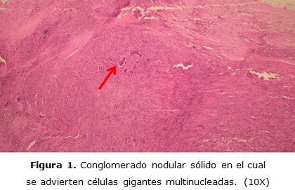
Results: a total of six cases of giant cell tumor of the tendon sheath were diagnosed, corresponding 50% to each sex; the majority were under 40 years old; and in all cases the site of presentation of the tumor was the hand; with a predominance in the palm region of the thumb. None of the cases manifested pain. In the morphological study, the characteristic histological elements of the giant cell tumor of the tendon sheath were observed microscopically.
Conclusions: the six patients studied presented the typical symptoms of the Giant cell tumor of the tendon sheath, referring to an increase in volume that was corroborated with the physical examination by palpating a mass of subcutaneous, non-painful, smooth, soft tissue with well-defined borders. In the same way, histopathologically, the characteristic morphological elements were corroborated, for which it can be established that there was a clinical-pathological correlation of 100%.
DeCS: GIANT CELL TUMOR OF TENDON SHEATH/diagnostic imaging; GIANT CELL TUMOR OF TENDON SHEATH/pathology; GIANT CELL TUMOR OF TENDON SHEATH/epidemiology; GIANT CELL TUMOR OF TENDON SHEATH /classification; MEDICAL RECORDS.
Downloads
References
1. Lifchez SD, Kelamis JA. Surgery of the hand and wrist. En: Brunicardi FC, editor. Schwartz’s principles of surgery. 10ma ed. Nueva York: McGraw Hill Education; 2014:1787-828.
2. Erosa MA, Cortés SA, López A. Tumor de células gigantes de la vaina tendinosa en la mano. Rev Esp Méd Quirúrg [Internet]. 2012 [citado 17 Feb 2021];17(2):146-49. Disponible en: https://www.redalyc.org/articulo.oa?id=47323278015
3. Zhang Y, Huang J, Ma X, Wang X, Zhang C, Chen L. Giant cell tumor of the tendon sheath in the foot and ankle: case series and review of the literature. J Foot Ankle Surg [Internet]. 2013 [citado 17 Feb 2021];52:24-7. Disponible en: https://www.jfas.org/article/S1067-2516(12)00425-5/pdf DOI:https://doi.org/10.1053/j.jfas.2012.09.008
4. Wang CS, Duan Q, Xue YJ, Huang XM, Wang LL, Chen ZY, et al. Giant cell tumor of tendon sheath with bone invasion in extremities: analysis of clinical and imaging findings. Radiol Med [Internet]. 2015 [citado 17 Feb 2021];120(8):745-52. Disponible en: https://www.ncbi.nlm.nih.gov/pubmed/25698301 DOI: 10.1007/s11547-015-0520-6
5. Rodríguez M, Ramos A, Domínguez MA, Salcedo DL. Tumor de células gigantes de la vaina tendinosa. Rev Cent Dermatol Pascua [Internet]. 2010 [citado 17 Feb 2021];19(3):108-11. Disponible en: https://www.medigraphic.com/pdfs/derma/cd-2010/cd103c.pdf
6. Jaffe HL, Lichtenstein L, Sutro CJ. Pigmented villonodular synovitis, bursitis and tenosynovitis. Arch Pathol. 1941;31:731-65.
7. Alarcón HH, Enríquez MJ, Novales SJ, Pérez CS. Tumor de células gigantes de la vaina tendinosa. Rev Cent Dermatol Pascua [Internet]. 2001 [citado 17 Feb 2021];10(2):90-3. Disponible en: https://www.medigraphic.com/pdfs/derma/cd-2001/cd012h.pdf
8. Ramos LR, Guerra OA, Casas P, Arias F. Giant cell tumor of the tendon sheaths of the fingers. Reumato Clínic [Internet]. 2015 [citado 17 Feb 2021];11:252-4. Disponible en: https://www.reumatologiaclinica.org/es-giant-cell-tumor-tendon-sheaths-articulo-S2173574314001774 DOI: 10.1016/j.reumae.2014.12.003
9. Mena E, Martín-Miramon JC, Berná L, Veintemillas M, Marín A, Valls R, et al. Tumor de células gigantes de la vaina tendinosa: hallazgos característicos de la gammagrafía ósea y correlación con la RMN. Rev Esp Med Nucl. 2009;28(5):249–52. DOI: 10.1016/j.remn.2009.04.002
10. Beekman F. Giant cell tumours of the tendon sheaths. Ann Surg [Internet]. 1915 [citado 17 Feb 2021];62:738-45. Disponible en:https://www.ncbi.nlm.nih.gov/pubmed/17863468 DOI:10.1097/00000658-191512000-00010
11. Wan JM, Magarelli N, Peh WC, Guglielmi G, Shek TW. Imaging of giant cell tumour of thetendonsheath. Radiol Med [Internet]. 2010 [citado 17 Feb 2021];115:141-51. Disponible en: https://www.ncbi.nlm.nih.gov/pubmed/20077044 DOI:10.1007/s11547-010-0515-2
12. Geschickter CF, Copeland MM. Tumors of bone. 3rd ed. Philadelphia: Lippincott; 1949:365-77.
13. Wright CJE. Benign giant-cell synovioma: an investigation of 85 cases. Br J Surg [Internet]. 1951 [citado 17 Feb 2021];38:257-71. Disponible en: https://onlinelibrary.wiley.com/doi/abs/10.1002/bjs.18003815102 DOI: 10.1002/bjs.18003815102
14. Heck RK. Benign tumors. En: Canale ST, Beaty JH, editors. Campbell’s Operative Orthopaedics. 11th ed. Maryland: Mosby; 2007:869-71.
15. Briët JP, Becker SJ, Oosterhoff TC, Ring D. Giant cell tumor of tendonsheath. Arch Bone Jt Surg [Internet]. 2015 [citado 17 Feb 2021];3:19-21. Disponible en: https://europepmc.org/abstract/med/25692164
16. Ho CY, Maleki Z. Giant cell tumor of tendon sheath: cytomorphologic and radiologic findings in 41 patients. Diagn Cytopathol [Internet]. 2012 [citado 17 Feb 2021];40:94-8. Disponible en: https://www.ncbi.nlm.nih.gov/pubmed/22454238 DOI:10.1002/dc.22840
17. Díaz JM, Domínguez Cherit J, López NS. Tumor de células gigantes de la vaina tendinosa: estudio retrospectivo de siete casos. Derma Cosm Quirúrg [Internet]. 2017 [citado 17 Feb 2021];15(1):11-3. Disponible en: https://www.medigraphic.com/pdfs/cosmetica/dcm-2017/dcm171c.pdf
18. Terada T. Giant cell tumor of the tendon sheath composed largely of epithelioidhistiocytes. Int J Clin Exp Pathol [Internet]. 2012 [citado 17 Feb 2021];5(4):374-76. Disponible en: http://www.ijcep.com/files/IJCEP1112002.pdf
19. Al Qattan MM. Giant cell tumor of tendon sheath: classification and recurrence rate. J Hand Surg Br [Internet]. 2001 [citado 17 Feb 2021];26(1):72-5. Disponible en: https://www.ncbi.nlm.nih.gov/pubmed/11162024 DOI:10.1054/jhsb.2000.0522
20. Fritchie KJ. Soft tissue and bone pathology. En: Reisner HM, editor. Pathology: a modern case study. Nueva York: McGraw Hill Education; 2015:483-526.
21. Tejera A, Ruiz I, González T, Solís E. Primary giant cell tumor of soft tissue in the finger. Dermatol Online J [Internet]. 2008 [citado 17 Feb 2021];14(6):7. Disponible en: https://www.ncbi.nlm.nih.gov/pubmed/18713588
Published
How to Cite
Issue
Section
License
Copyright (c) 2021 Alberto José Alberto Piamo-Morales, Daisy Ferrer-Marrero, Isnerio Arzuaga-Anderson, Digna Chávez-Jiménez, Lourdes Palma-Machado

This work is licensed under a Creative Commons Attribution-NonCommercial 4.0 International License.
Copyright: Camagüey Medical Archive Magazine, offers immediately after being indexed in the SciELO Project; Open access to the full text of the articles under the principle of making available and free the research to promote the exchange of global knowledge and contribute to a greater extension, publication, evaluation and extensive use of the articles that can be used without purpose As long as reference is made to the primary source.
Conflicts of interest: authors must declare in a mandatory manner the presence or not of conflicts of interest in relation to the investigation presented.
(Download Statement of potential conflicts of interest)
The Revista Archivo Médico de Camagüey is under a License Creative Commons Attribution-Noncommercial-No Derivative Works 4.0 International (CC BY 4.0).
This license allows others to distribute, to mix, to adjust and to build from its work, even for commercial purposes, as long as it is recognized the authorship of the original creation. This is the most helpful license offered. Recommended for maximum dissemination and use of licensed materials. The full license can be found at: https://creativecommons.org/licenses/


 22 julio 2025