

Common pattern and anatomical variants of the anterior portion of the arterial circle of the brain
Keywords:
círculo arterial del cerebro; variantes anatómicas; patrón común; arteria cerebral anterior; arteria comunicante anterior.Abstract
Background: the brain is an organ that requires an uninterrupted supply of blood that is guaranteed by the circle of Willis.
Objective: to describe the common pattern and anatomical variants of the anterior portion of the circle of Willis.
Methods: a cross-sectional descriptive study was conducted in the department of Morphological Sciences at the University of Medical Sciences in Camagüey from December 2014 until May 2016. The universe was composed of 50 brains, which were washed with common water, and fixed in a 10 % formalin solution. Then a macroscopic dissection was performed. The variables studied were: origin, number, path and external diameter. Data were processed using the SPSS statistical package for Windows version 21.0, and descriptive statistics were applied. The results were presented in tables and images.
Results: the origin of the precommunicating segment of the anterior cerebral artery from the internal carotid artery, forward and inward to the interhemispheric fissure, and join at the one on both sides in front of the optic nerve through the anterior communicating artery, with variation in the path and in the external diameter, without numerical variants: the anterior communicating artery with variations in the path, number and external diameter.
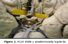
Conclusions: the common pattern and anatomical variants of the anterior portion of the arterial circle of the brain were presented in equal proportion. The most observed anatomical variant was the duplication of the anterior communicating artery.
DeCS: ANTERIOR CEREBRAL ARTERY; CIRCLE OF WILLI; ANATOMIC VARIATION; DISSECTION; EPIDEMIOLOGY, DESCRIPTIVE.
Downloads
References
1.Lippert H. Anatomía. 4ta ed. Madrid: España; 2000.
2.Arráez Aybar LA, Navia Alvarez P, Fuentes-Redondo T, Bueno-López JL. Thomas Willis, a pioneer in translational research in anatomy (on the 350th anniversary of Cerebri anatome). J Anat [Internet]. 2015 [citado 2016 May 29];226(3):[about 12 p.]. Available from: http://onlinelibrary.wiley.com/store/10.1111/joa.12273/asset/joa12273.pdf?v=1&t=itq5lj48&s=a528aca83e843a912b3ce5ce1c22b0c3fb1e3fde
3.Latarjet M, Ruiz Liard A. Anatomía Humana. 2a ed. México: Editorial Médica panamericana; 1990.
4.Siddiqi H, Tahir M, Lone K. Variations in cerebral arterial circle of Willis in adult Pakistani population. J Coll Physicians Surg Pak [Internet]. 2013 [citado 2016 May 9];23(9):[about 5 p.]. Available from: http://applications.emro.who.int/imemrf/J_Coll_Physicians_Surg_Pak/J_Coll_Physicians_Surg_Pak_2013_23_9_615_619.pdf
5.Moore KL, Dalley AF. Clinically oriented anatomy. 4ta ed. Philadelphia: Lippincott Williams Wilkins; 1999.
6.Zhou H, Sun J, Ji X, Lin J, Tang S, Zeng J, et al. Correlation Between the Integrity of the Circle of Willis and the Severity of Initial Noncardiac Cerebral Infarction and Clinical Prognosis. Medicine [Internet]. 2016 [citado 2017 Feb 21]; [about 7 p.]. Available from:
https://www.ncbi.nlm.nih.gov/pmc/articles/PMC4998866/pdf/medi-95-e02892.pdf
7.Monroy Sosa A, Pérez Cruz J, Reyes Soto G, Delgado Hernández C, Macías Duvignau M, Delgado Reyes L. Importancia de la anatomía microquirúrgica del complejo A1-arteria comunicante anterior. Cir Cir [Internet]. 2013 [citado 2016 May 9];81(4)[aprox. 7 p.]. Disponible en: http://www.redalyc.org/html/662/66228318003/.
8.Ferré J, Niederberger E, Morandi X, Raoult H, Carsin Nicol B, Gauvrit J. Anatomical variations of the anterior cerebral arterial circle visualized by multidetector computed tomography angiography: Comparison with 3D rotational angiography. J neuroradiol [Internet]. 2013 [citado 2014 Feb 19];[about 9 p.]. Available from: http://posterng.netkey.at/esr/viewing/index.php?module=viewing_poster&task=viewsection&pi=105916&ti=336123&searchkey
9.Kedia S, Daisy S, Mukherjee K, Salunke P, Srinivasa R, Narain M. Microsurgical anatomy of the anterior cerebral artery in Indian cadavers. Neurology India [Internet]. 2013 [citado 9 May 2016];61(2):[aprox. 4 p.]. Available from: http://www.neurologyindia.com/article.asp?issn=00283886;year=2013;volume=61;issue=2;spage=117;epage=121;aulast=Kedia
10.Pino Mederos Y. La norma anatómica del sistema de la Arteria Cerebral Anterior en el hombre adulto [tesis]. Camagüey: Universidad de Ciencias Médicas; 2006.
11.Yasargil MG. Microneurosurgery. New York: Thieme Stratton; 1984.
12.Madrid Muñiz C, Arias Ortega M, Cortes Vela JJ, Garcia Nieto JC, Valentín Martín AB. Gonzalez-Spinola SG, et al. Estudio de las variantes del Polígono de Willis [Internet]. España: Sociedad Española de Radiología Médica (SERAM); 2014 [citado 9 May 2016]. Disponible en: http://pdf.posterng.netkey.at/download/index.php?module=get_pdf_by_id&poster_id=123827
13.Rhoton AL. Thesupratentorialarteries. Neurosurgery. 2002;51(1):82-05.
14.González X; Landó F. Angiotomografía Cerebral: variantes anatómicas más frecuentes del Polígono de Willis. Ensayo Iconográfico [Internet]. Uruguay: IX Congreso Uruguayo de Radiología; 2014 [citado 9 May 2016]. Disponible en: http://webcir.org/revistavirtual/articulos/noviembre14/uruguay/poligono_de_willis_esp.pdf
15.Martínez F, Espagnuolo E, Calvo Rubal A, Laza S, Sgarbi N, Soria Vargas VR, et al. Variaciones del sector anterior del Polígono de Willis. Correlación anatomo-angiográfica y su implicancia en la cirugía de aneurismas intracraneanos. Neurocirugía [Internet]. 2016 [citado 26 May 2016];15:[aprox. 10 p.]. Disponible en:
http://www.revistaneurocirugia.com/es/variacionesdelsectoranteriordel/articulo/S1130147304704492/
16.Raghavendra K, Shirol VS, Dixit D, Reddy AK, Desai SP. Circle of Willis andits variations; morphometric study in adult human cadavers. Int J Med Res Health Sci [Internet]. 2014 [citado 2016 May 26];3(2):[about 6 p.]. Available from: http://ijmrhs.com/vi32/32%20Raghavendra%20etal.pdf
17.Hernández Luna J, Casares Cruz K, Rendón Macías R, Licea Medina D, Castillo Lima J. Evaluación con angiorresonancia magnética nuclear de las variantes anatómicas del círculo arterial cerebral. An Radiol Mex [Internet]. 2015 [citado 9 May 2016];14(3):[aprox. 6 p.]. Disponible en: http://web.a.ebscohost.com/ehost/pdfviewer/pdfviewer?vid=24&sid=bd518b17-e153-4cf0-b57d-3473535aa667%40sessionmgr4007&hid=4114
18.Klimek Piotrowska W, Kopeć M, Kochana M, Krzyżewski R, Tomaszewski K, Walocha J, et al. Configurations of the circle of Willis: a computed tomography angiography based study on a Polish population. Folia Morphol [Internet]. 2013 [citado 2016 May 9];72(4):[about 3 p.]. Available from: https://journals.viamedica.pl/folia_morphologica/article/viewFile/FM.2013.0049/26223.pdf
19.Hannequin P, Peltier J, Destrieux C, Velut S, Havet E, Le Gars D. The inter-optic course of a unique precommunicating anterior cerebral artery with aberrant origin of an ophthalmic artery: an anatomic case report. Surg Radiol Anat [Internet]. 2013 [citado 2016 May 26];35(3):[about 2 p.]. Available from: http://web.a.ebscohost.com/ehost/pdfviewer/pdfviewer?vid=3&sid=7bd7e6b6-e00b-499e-9040-953bfc464d95%40sessionmgr4009&hid=4114
20.Okano N, Uchino A, Saito N, MaruyamaH. Left carotid-anterior cerebral artery anastomosis diagnosed by MR angiography: a case report. Surgradiolanat [Internet]. 2015 [citado 2016 May 19];37:[about 2 p.]. Available from:
https://link.springer.com/article/10.1007%2Fs00276-014-1358-7
21.Qiu C, Zhang Y, Xue C, Jiang S, Zhang W. MRA study on variation of the circle of Willis in healthy Chinese male adults. Biomed Res Int[Internet]. 2015[citado 2016 May 27]:[about 3 p.]. Available from: https://www.hindawi.com/journals/bmri/2015/976340/.
22.Krzyżewski R, Tomaszewski K, Kochana M, Kopeć M, Klimek-Piotrowska W, Walocha J. Anatomical variations of the anterior communicating artery complex: gender relationship. Surg Radiol Anat [Internet]. 2015 [citado 2016 May 9];37:[about 5 p.]. Available from: http://fmc.czasopisma.pan.pl/images/data/fmc/wydania/No_1_2014/Krzyewski.pdf
23.Papantchev V, Stoinova V, Aleksandrov A, Todorova Papantcheva D, Hristov S, Ovtscharoff W, et al. The role of Willis circle variations during unilateral selective cerebral perfusion: a study of 500 circles. Eur J Cardiothorac Surg [Internet]. 2013 [citado 2016 May 26];44(4):[about 10 p.]. Available from: https://www.researchgate.net/publication/235885471_The_role_of_Willis_circle
24.Huang J, Germanwala AV, Tamargo RJ. Anterior Communicating Artery Aneurysms. En: Winn HR, editor. Youmans Neurological Surgery. 6th ed [Internet]. Madrid: Elsevier Saunders; 2011 [citado 2016 Jun 2]. Available from: https://www.clinicalkey.es/#!/content/book/3-s2.0-B9781416053163003713
25.Choi JH, Jo K, Kim KH, Jeon P, Yeon JY, Kim JS, et al. Morphological risk factors for the rupture of anterior communicating artery aneurysms: the significance of fenestration. Neuroradiology [Internet]. 2016 [citado 2016 Jun 2];58(2):[about 5 p.]. Available from: http://web.a.ebscohost.com/ehost/pdfviewer/pdfviewer?vid=18&sid=bd518b17-e153-4cf0-b57d-3473535aa667%40sessionmgr4007&hid=4114
Published
How to Cite
Issue
Section
License
Copyright (c) 2017 Mayrelis Pacheco Mayedo, Mayda Estrella Durán Matos, Olga Lidia Cuba Yordi, Luisa Serrano González, Yanil Rosales Almeida, Johnny Loret de Mola Nicolau

This work is licensed under a Creative Commons Attribution-NonCommercial 4.0 International License.
Copyright: Camagüey Medical Archive Magazine, offers immediately after being indexed in the SciELO Project; Open access to the full text of the articles under the principle of making available and free the research to promote the exchange of global knowledge and contribute to a greater extension, publication, evaluation and extensive use of the articles that can be used without purpose As long as reference is made to the primary source.
Conflicts of interest: authors must declare in a mandatory manner the presence or not of conflicts of interest in relation to the investigation presented.
(Download Statement of potential conflicts of interest)
The Revista Archivo Médico de Camagüey is under a License Creative Commons Attribution-Noncommercial-No Derivative Works 4.0 International (CC BY 4.0).
This license allows others to distribute, to mix, to adjust and to build from its work, even for commercial purposes, as long as it is recognized the authorship of the original creation. This is the most helpful license offered. Recommended for maximum dissemination and use of licensed materials. The full license can be found at: https://creativecommons.org/licenses/


 22 julio 2025