

Lesiones del tendón poplíteo
Resumen
Introducción: Las estructuras que conforman la esquina posterolateral de la rodilla sufren de afecciones tanto traumáticas como degenerativas, entre ellas se encuentran las lesiones del tendón poplíteo.
Objetivo: Actualizar y brindar información sobre las lesiones aisladas y combinadas del tendón poplíteo.
Métodos: La búsqueda y análisis de la información se realizó en un periodo de 61 días (primero de agosto al 30 de septiembre de 2023), se emplearon las siguientes palabras: popliteus tendon injury, posterolateral complex, ligament injury AND knee, tendon injury AND knee. A partir de la información obtenida se realizó una revisión bibliográfica de un total de 153 artículos publicados en las bases de datos PubMed, HINARI, SciELO, Researchgate, Ebsco, Scopus, Medscape y Medline mediante el gestor de búsqueda y administrador de referencias EndNote, de ellos, se utilizaron 30 citas seleccionadas para realizar la revisión.
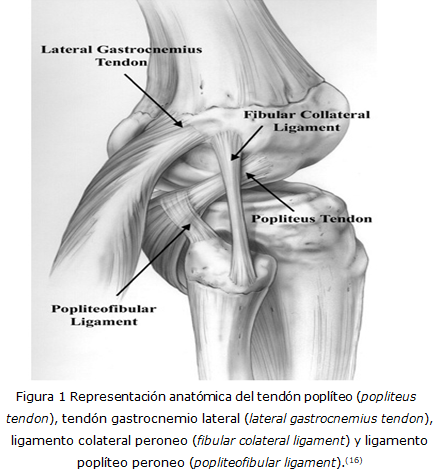
Resultados: Se hizo referencia a la anatomía esencial del tendón poplíteo, así como su relación con las estructuras de la esquina posterolateral. Se abordó el mecanismo de producción, clasificación, aspectos clínicos y estudios imagenológicos de las lesiones de esta zona. En relación al tratamiento, se describieron las modalidades y sus indicaciones.
Conclusiones: Las lesiones del tendón poplíteo pueden presentarse de forma aislada o combinada tanto por lesiones traumáticas como degenerativas. Para el diagnóstico de la enfermedad se necesitan tres pilares fundamentales: el clínico, imagenológico y artroscópico. La modalidad de tratamiento depende si se trata de una lesión aislada o combinada.
DeCS: TRAUMATISMOS DE LA RODILLA; TENDINOPATÍA/clasificación; TENDINOPATÍA/cirugía; ARTROSCOPÍA; REVISIÓN.
Descargas
Citas
1. Algazwi DAR, Tian QS, Elizabeth GL, Ellis ES, Teng VSY, Hallinan JTPD. Isolated popliteus tendon avulsion fracture. Am J Phys Med Rehabil [Internet]. 2019 [citado 30 Ago 2023];98(11):e140-e141. Disponible en: https://doi.org/10.1097/PHM.0000000000001181
2. Maniar AR, White AE, Musahl V, Ranawat A. Posterolateral corner of the knee: an update on current evaluation and management strategies. J Am Acad Orthop Surg [Internet]. 2023 [citado 30 Ago 2023]. Disponible en: https://doi.org/10.5435/JAAOS-D-23-00278
3. Aumann EK, Aksu T, Atansay V, Kara AN, Aksu N. Relationship of popliteus sulcus depth and tibiofemoral rotational alignment with popliteus tendinitis in professional folk dancers exposed to turnout positions: an MRI analysis. Med Probl Perform Art [Internet]. 2019 [citado 30 Ago 2023];34(3):141-146. Disponible en: https://doi.org/10.21091/mppa.2019.3024
4. Mutou M, Abe Y, Kataoka H, Fuzisawa T, Takahashi Y. Anatomical reconstruction of the posterolateral corner of the knee preserving dynamic function of the popliteus tendon complex. Asia Pac J Sports Med Arthrosc Rehabil Technol [Internet]. 2022 Mar [citado 30 Ago 2023];28:1-5. Disponible en: https://www.sciencedirect.com/science/article/pii/S2214687322000024?via%3Dihub
5. Blackwell C, Selley R, Taber CE, Benitez CL, Marx RG. Chronic popliteus tendon avulsion fracture with chronic knee pain and locking: a case report. JBJS Case Connect [Internet]. 2022 [citado 30 Ago 2023];12(1). Disponible en: https://doi.org/10.2106/JBJS.CC.21.00477
6. Zabrzynski J, Huri G, Yataganbaba A, Paczesny L, Szwedowski D, Zabrzynska A, et al. Current concepts on the morphology of popliteus tendon and its clinical implications. Folia Morphol (Warsz) [Internet]. 2021 [citado 30 Ago 2023];80(3):505-513. Disponible en: https://doi.org/10.5603/FM.a2020.0106
7. Farrell C, Kiel J, Seemann L, Pujalte GGA. Popliteus tendon injuries. Orthopedics [Internet]. 2023 [citado 30 Ago 2023];46(4):e193-e198. Disponible en: https://doi.org/10.3928/01477447-20220719-10
8. Morrissey CD, Knapik DM. Prevalence, mechanisms, and return to sport after isolated popliteus injuries in athletes: a systematic review. Orthop J Sports Med [Internet]. 2022 [citado 30 Ago 2023];10(2):[aprox. 3 p.]. Disponible en: https://doi.org/10.1177/23259671211073617
9. Azzopardi C, Kiernan G, Botchu R. Imaging review of normal anatomy and pathological conditions involving the popliteus. J Clin Orthop Traum [Internet]. 2021 [Citado 30 Ago 2023];18:224-229. Disponible en: https://www.ncbi.nlm.nih.gov/pmc/articles/PMC8173307/
10. Sanchez Munoz E, Lozano Hernanz B, Zijl JAC, Passarelli Tirico LE, Angelini FJ, Verdonk PCM, et al. Accuracy of magnetic resonance imaging in the diagnosis of multiple ligament knee injuries: a multicenter study of 178 patients. Am J Sports Med [Internet]. 2023 [citado 30 Ago 2023];51(2):429-436. Disponible en: https://journals.sagepub.com/doi/10.1177/03635465221145697
11. Koong DP, An VVG, Lorentzos P, Moussa P, Sivakumar BS. Non-operative rehabilitation of isolated popliteus tendon rupture in a Rugby player. Knee Surg Relat Res [Internet]. 2018 [citado 30 Ago 2023];30(3):269-272. Disponible en: https://doi.org/10.5792/ksrr.17.072
12. Faraco Sobrado M, Partezani Helito C, Ponte Melo L, Marangoni Asperti A, Gomes Gobbi R, Janson Angelini F. Anatomical study of the posterolateral ligament complex of the knee: LCL and popliteus tendon. Acta Ortop Bras [Internet]. 2021 [citado 30 Ago 2023];29(5):249-252. Disponible en: http://doi.org/10.1590/1413-785220212905241252
13. Porrino J, Sharp JW, Ashimolowo T, Dunham G. An Update and comprehensive review of the posterolateral corner of the knee. Radiol Clin North Am [Internet]. 2018 Nov [citado 30 Ago 2023];56(6):935-951. Disponible en: https://doi.org/10.1016/j.rcl.2018.06.006
14. Weiss S, Krause M, Frosch KH. Posterolateral corner of the knee: a systematic literature review of current concepts of arthroscopic reconstruction. Arch Orthop Trauma Surg [Internet]. 2020 [citado 30 Ago 2023];140(12):2003-2012. Disponible en: https://doi.org/10.1007/s00402-020-03607-z
15. Wood A, Boren M, Dodgen T, Wagner R, Patterson RM. Muscular architecture of the popliteus muscle and the basic science implications. Knee [Internet]. 2020 [citado 30 Ago 2023];27(2):308-314. Disponible en: https://doi.org/10.1016/j.knee.2019.12.001
16. Swinford S, La Prade R, Engebresten L, Cohen M, Safran M. Biomechanics and physical examination of the posteromedial and posterolateral knee: state of the art. J ISAKOS [Internet]. 2020 [citado 30 Ago 2023];5:378-388. Disponible en: https://doi.org/10.1136/jisakos-2018-000221
17. Annear P, Arora M. Isolated popliteal tendon avulsion: current understanding and approach to management. J Arthrosc Joint Surg [Internet]. 2018 [citado 30 Ago 2023];5(3):145-148. Disponible en: http://doi.org/10.1016/j.jajs.2018.02.004
18. Wong KC, Mohamad N, Md Yusoff BAH. Popliteus tendon injury: a rare cause of acute locked knee. Cureus [Internet]. 2023 [citado 30 Ago 2023];15(5):e38655. Disponible en: https://doi.org/10.7759/cureus.38655
19. Rodriguez AN, Liechti DJ, La Prade RF. Open popliteus tendon reconstruction using a hamstring tendon autograft. Arthrosc Tech [Internet]. 2023 [citado 30 Ago 2023];12(4):e453-e457. Disponible en: https://doi.org/10.1016/j.eats.2022.11.028
20. Yoo HJ, Ryu KN, Park JS, Jin W, Park SY, Kang HJ, et al. Preoperative meniscus: pitfalls and traps to avoid. Taehan Yongsang Uihakhoe Chi [Internet]. 2022 [citado 30 Ago 2023];83(3):582-596. Disponible en: https://doi.org/10.3348/jksr.2021.0002
21. Al Dosari M, Elmhiregh A, Hammad M, Alam S, Hameed S. Rare presentation of lateral meniscus tear with pathognomonic MRI finding. Int J Surg Case Rep [Internet]. 2019 [citado 30 Ago 2023];65:339-343. Disponible en: https://doi.org/10.1016/j.ijscr.2019.11.025
22. Grassi A, Pizza N, Andrea Lucidi G, Macchiarola L, Mosca M, Zaffagnini S. Anatomy, magnetic resonance and arthroscopy of the popliteal hiatus of the knee: normal aspect and pathological conditions. EFORT Open Rev [Internet]. 2021 [citado 30 Ago 2023];6(1):61-74. Disponible en: https://doi.org/10.1302/2058-5241.6.200089
23. Kompel A, Haran PH, Murakami AM, Engebretsen L, Jarraya M, Roemer F, et al. MRI-detected knee ligament sprains and associated internal derangement in athletes competing at the Rio de Janeiro 2016 summer Olympics. Open Access J Sports Med [Internet]. 2021 [citado 30 Ago 2023];12:23-32. Disponible en: https://doi.org/10.2147/OAJSM.S292763
24. Saini NK, Yadav S, Jain VK, Shukla A. Measurement of distance between femoral insertion of fibular collateral ligament and popliteus tendon: a magnetic resonance imaging based study. J Clin Orthop Trauma [Internet]. 2021 [citado 30 Ago 2023];17:139-142. Disponible en: https://doi.org/10.1016/j.jcot.2021.02.023
25. Mishra P, Goyal A, Topgia C, Lal H, Kumar S, Ajay A. Measurement of distance between femoral insertion of fibular collateral ligament and popliteus: a cadaveric study in Indian population. Indian J Orthop [Internet]. 2022 [citado 30 Ago 2023];56(10):1717-1721. Disponible en: https://doi.org/10.1007/s43465-022-00711-7
26. Newcomb NL, Kenneally CM, Yerdon HN, Barry PA. Diving for the basketball: an isolated popliteus rupture in an adolescent female with 6 year follow-up. Glob Pediatr Health [Internet]. 2021 [citado 30 Ago 2023];8. Disponible en: https://doi.org/10.1177/2333794X211020248
27. Ricci V, Özçakar L. Ultrasound imaging for lateral knee pain: popliteus tendon highlighted. Med Ultrason [Internet]. 2018 [citado 30 Ago 2023];20(3):403-404. Disponible en: https://doi.org/10.11152/mu-1616
28. Koukoulias NE, Dimitriadis T, Boutovinos AP, Germanou E. Isolated popliteus tendon avulsion: fully arthroscopic repair with suture anchor: a case report. JBJS Case Connect [Internet]. 2020 [citado 30 Ago 2023];10(3):e20.00159. Disponible en: https://doi.org/10.2106/JBJS.CC.20.00159
29. Zappia M, Reginelli A, Chianca V, Carfora M, Di Pietto F, Iannella G, et al. MRI of popliteo-meniscal fasciculi of the knee: a pictorial review. Acta Biomed [Internet]. 2018 [citado 30 Ago 2023];89:suppl 1:7-17. Disponible en: https://doi.org/10.23750/abm.v89i1-S.7007
30. Arner JW, Johannsen AM, Ruzbarsky JJ, Godin JA. Open popliteal tendon repair. Arthrosc Tech [Internet]. 2021 [citado 30 Ago 2023];10(2):e499-e505. Disponible en: https://doi.org/10.1016/j.eats.2020.10.031
Publicado
Cómo citar
Número
Sección
Licencia
Derechos de autor 2024 Alejandro Alvarez-López, Valentina Valdebenito-Aceitón, Sergio Ricardo Soto-Carrasco, Tuan Nguyen-Pham

Esta obra está bajo una licencia internacional Creative Commons Atribución-NoComercial 4.0.
La Revista Archivo Medico Camagüey, ofrece de forma inmediata después de ser indexada en el Proyecto SciELO; acceso abierto al texto completo de los artículos bajo el principio de hacer disponible y gratuita la investigación para favorecer el intercambio del conocimiento global y coadyuvar a una mayor extensión, publicación, evaluación y uso extensivo de los artículos que se exponen pudiendo ser utilizados, sin fines comerciales, siempre y cuando se haga referencia a la fuente primaria.
Carta De Declaración De Autoría u Derechos De Autor(a)
Conflictos de intereses: los autores deberán declarar de forma obligatoria la presencia o no de conflictos de intereses en relación con la investigación presentada. (Descargar Plantilla para declarar confictos de intereses)
La Revista Archivo Médico Camagüey se encuentra bajo una
Licencia Creative Commons Reconocimiento-NoComercial 4.0 International (CC BY NC 4.0).
Esta licencia permite a otros distribuir, mezclar, ajustar y construir a partir de su obra, incluso con fines comerciales, siempre que le sea reconocida la autoría de la creación original. Esta es la licencia más servicial de las ofrecidas. Recomendada para una máxima difusión y utilización de los materiales sujetos a la licencia. La licencia completa puede consultarse en: https://creativecommons.org/licenses/


