Artroscopia y fibulectomía parcial simultánea en pacientes con gonartrosis y deformidad en varo

Resumen
Fundamento: la gonartrosis es una enfermedad frecuente relacionada con el incremento de la calidad y expectativa de vida de la población, en la evolución de este padecimiento existen factores que aceleran sus manifestaciones entre ellos la deformidad en varo.
Objetivo: evaluar los resultados de la técnica quirúrgica combinada de artroscopia, fibulectomía parcial y proximal en pacientes con gonartrosis y deformidad en varo.
Métodos: se realizó un estudio cuasi experimental modalidad antes y después sin grupo de control en 25 pacientes con el diagnóstico de gonartrosis primaria asociada a deformidad en varo, en el Hospital Universitario Manuel Ascunce Domenech de la provincia Camagüey desde abril 2016 a agosto de 2019. La investigación tiene un nivel de evidencia II recomendación B.
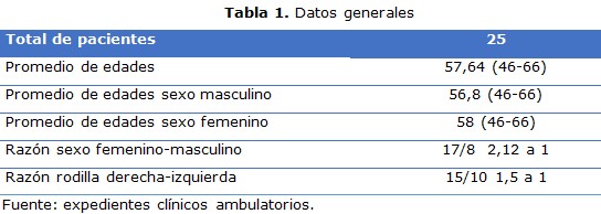
Resultados: predominio del sexo femenino al masculino con una razón de 2,12 a 1. La enfermedad intrarticular más frecuente fue la lesión de meniscos y cartílagos grados III/IV. Se encontró significación entre un antes y después al aplicar las escalas evaluativas. El procedimiento artroscópico más empleado fue la meniscectomía.
Conclusiones: la realización simultanea de artroscopia y fibulectomía parcial proximal es una técnica efectiva y sencilla con un mínimo de complicaciones, permite corregir la deformidad angular de la extremidad, al mismo tiempo de tratar lesiones intrarticulares, en especial las de menisco y cartílago.
DeCS: ARTROSCOPÍA/métodos; GENU VARUM/cirugía; OSTEOARTRITIS DE LA RODILLA/cirugía; MENISCECTOMÍA; PERONÉ/cirugía.
Palabras clave
Referencias
Klatt BA, Chen A, Tuan RT. Arthritis and other cartilage disorders. En: Cannada LK. OKU 11. Rosemont: Am Acad Orthop Surg; 2014.p.207-22.
Bennell KL, Dobson F, Roos EM, Skou ST, Hodges P, Wrigley TV, et al. Influence of biomechanical characteristics on pain and function outcomes from exercise in medial knee osteoarthritis and varus malalignment: exploratory analyses from a randomized controlled trial. Arthritis Care Res (Hoboken). 2015 Sep;67(9):1281-8.
Dell' Isola A, Allan R, Smith SL, Marreiros SS, Steultjens M. Identification of clinical phenotypes in knee osteoarthritis: a systematic review of the literature. BMC Musculoskelet Disord. 2016 Oct;17(1):425.
van Tunen JAC, Dell' Isola A, Juhl C, Dekker J, Steultjens M, Thorlund JB, et al. Association of malalignment, muscular dysfunction, proprioception, laxity and abnormal joint loading with tibiofemoral knee osteoarthritis - a systematic review and meta-analysis. BMC Musculoskelet Disord. 2018 Jul;19(1):273.
Bastick AN, Belo JN, Runhaar J, Bierma-Zeinstra SM. What are the prognostic factors for radiographic progression of knee osteoarthritis? A meta-analysis. Clin Orthop Relat Res. 2015 Sep;473(9):2969-89.
Baier C, Benditz A, Koeck F, Keshmiri A, Grifka J, Maderbacher G. Different kinematics of knees with varus and valgus deformities. J Knee Surg. 2018 Mar;31(3):264-9.
Driban JB, Mc Alindon TE, Amin M, Price LL, Eaton CB, Davis JE, et al. Risk factors can classify individuals who develop accelerated knee osteoarthritis: data from the osteoarthritis initiative. J Orthop Res. 2018 Mar;36(3):876-80.
Rothental PB. Knee osteoarthritis. En: Scott WN. Insall&Scott Surgery of the Knee. 6th ed. Philadelphia: Elsevier;2018.p.992-7.
Ji W, Luo C, Zhan Y, Xie X, He Q, Zhang B. A residual intra-articular varus after medial opening wedge high tibial osteotomy (HTO) for varus osteoarthritis of the knee. Arch Orthop Trauma Surg. 2019 Jun;139(6):743-50.
Stickley CD, Presuto MM, Radzak KN, Bourbeau CM, Hetzler RK. Dynamic varus and the development of Iliotibial Band Syndrome. J Athl Train. 2018 Feb;53(2):128-34.
Wang X, Wei L, Lv Z, Zhao B, Duan Z, Wu W, et al. Proximal fibular osteotomy: a new surgery for pain relief and improvement of joint function in patiets with knee osteoarthritis. J Inter Medical Research. 2017 Jan;45(1):282-9.
Yazdi H, Mallakzadeh M, Mohtajeb M, Farshidfar SS, Bagherty A, Givehchian B. The effect of partial fibulectomy on contact pressure of the knee: a cadaveric study. Eur J Orthop Surg Traumatol. 2014 Oct;24(7):1285-9.
Vandekerckhove PTK, Matlovich N, Teeter MG, MacDonald SJ, Howard JL, Lantin BA. The relationship between constitutional alignment and varus osteoarthritis of the knee. Knee Surg Sports Traumatol Arthrosc. 2017 Sep;25(9):2873-9.
Hochberg MC, Altman RD, Brandt KD, Clark BM, Dieppe PA, Griffin MR, et al. Guidelines for the medical management of osteoarthritis. Part II: Osteoarthritis of the knee. Arthritis Rheum. 1995 Nov;38(11):1541-6.
Mochizuki T, Tanifuji O, Koga Y, Sato T, Kobayashi K, Nishino K, et al. Sex differences in femoral deformity determined using three-dimensional assessment for osteoarthritic knees. Knee Surg Sports Traumatol Arthrosc. 2017 Feb;25(2):468-76.
Omori G, Narumi K, Nishino K, Nawata A, Watanabe H, Tanaka M, et al. Association of mechanical factors with medial knee osteoarthritis: a cross-sectional study from Matsudai Knee Osteoarthritis Survey. J Orthop Sci. 2016 Jul;21(4):463-8.
Sharma L, Chmiel JS, Almagor O, Moisio K, Chang AH, Belisle L, et al. Knee instability and basic and advanced function decline in knee osteoarthritis. Arthritis Care Res (Hoboken). 2015 Aug;67(8):1095-102.
Morin V, Pailhé R, Duval BR, Mader R, Cognault J, Rouchy RC, et al. Gait analysis following medial opening-wedge high tibial osteotomy. Knee Surg Sports Traumatol Arthrosc. 2018 Jun;26(6):1838-1844.
Im GI, Kim MK, Lee SH. Relationship between knee alignment and radiographic markers of osteoarthritis: a cross-sectional study from a Korean population. Int J Rheum Dis. 2016 Feb;19(2):178-83.
Faschingbauer M, Renner L, Waldstein W, Boettner F. Are lateral compartment osteophytes a predictor for lateral cartilage damage in varus osteoarthritic knees? data from the Osteoarthritis Initiative. Bone Joint J. 2015 Dec;97-B(12):1634-9.
Cho SD, Youm YS, Kim JH, Cho HY, Kim KH. Patterns and influencing factors of medial meniscus tears in varus knee osteoarthritis. Knee Surg Relat Res. 2016 Jun;28(2):142-6.
Freisinger GM, Schmitt LC, Wanamaker AB, Siston RA, Chaudhari AMW. Tibiofemoral osteoarthritis and varus-valgus laxity. J Knee Surg. 2017 Jun;30(5):440-51.
Kelly JD. Meniscal injuries. New York: Springer; 2014.
Puthumanapully PK, Harris SJ, Leong A, Cobb JP, Amis AA, Jeffers J. A morphometric study of normal and varus knees. Knee Surg Sports Traumatol Arthrosc. 2014 Dec;22(12):2891-9.
Whelton C, Thomas A, Elson DW, Metcalfe A, Forrest S, Wilson C, et al. Combined effect of toe out gait and high tibial osteotomy on knee adduction moment in patients with varus knee deformity. Clin Biomech (Bristol, Avon). 2017 Mar;43:109-14.
Rezaeian ZS, Smith MM, Skaife TL, Harvey WF, Gross KD, Hunter DJ. Does knee malalignment predict the efficacy of realignment therapy for patients with knee osteoarthritis? Int J Rheum Dis. 2017 Oct;20(10):1403-12.
Iranpour Boroujeni T, Li J, Lynch JA, Nevitt M, Duryea M. A new method to measure anatomic knee alignment for large studies of OA: data from the Osteoarthritis Initiative. Osteoarthritis Cartilage. 2014 Oct;22(10):1668-74.
O'Connell M, Farrokhi S, Fitzgerald GK. The role of knee joint moments and knee impairments on self-reported knee pain during gait in patients with knee osteoarthritis. Clin Biomech (Bristol, Avon). 2016 Jan;31:40-6.
Qin D, Chen W, Wang J, Lv H, Ma W, Dong T, et al. Mechanism and influencing factors of proximal fibular osteotomy for treatment of medial compartment knee osteoarthritis: a prospective study. J Int Med Res. 2018 Aug;46(8):3114-23.
Yang ZY, Chen W, Li CX, Wang J, Shao DC, Hou ZY, et al. Medial compartment decompression by fibular osteotomy to treat medial compartment knee osteoarthritis: a pilot study. Orthopedics. 2015 Dec;38(12):e1110-4.
Enlaces refback
- No hay ningún enlace refback.

Esta obra está bajo una licencia de Creative Commons Reconocimiento-NoComercial-CompartirIgual 4.0 Internacional.


 La Revista está: Certificada por el CITMA
 Acreditados como: "Web de Interés Sanitario"
