

Nuclear Morphometric characterization of the healthy epidermis in patients with different ages and sex
Abstract
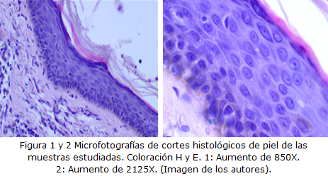
Introduction: The skin is a complex organ highly vulnerable to aging, a phenomenon that biologically causes changes at the tissue and cellular level. Usually the histological elements that characterize it are described with a qualitative approach, without taken age into account, however from the quantitative point of view, this aspect that morphometry makes possible has not been addressed in all its potentialities.
Objective: To characterize the behavior of morphometric indicators such as perimeter, area, nuclear volume in the cells of the spinous layer of the healthy epidermis, according to age and sex.
Methods: A case series study was carried out with 12 patients diagnosed with histopathology of basal cell carcinoma treated at the Oncology Center of the Provincial University Hospital Vladimir Ilich Lenin of Holguin, in the year 2019 and to which the tumor was removed by means of an excisional biopsy that included the lesion and a broad border of healthy skin. Theoretical and empirical methods were used, the latter based on techniques morphometric, then statistical analysis of the data obtained was performed and were reflected in tables.
Results: As age advances the nuclear perimeter, area, and volume decreased in both sexes.
Conclusions: Both the perimeter, the area and the nuclear volume decrease in the cell of the spinous layer of the healthy epidermis in both sexes as age advances, which translates into a decrease in nuclear size.
DeCS: EPIDERMIS; SKIN; SKIN AGING; CARCINOMA, BASAL CELL/pathology; CARCINOMA, BASAL CELL/therapy.
Downloads
References
1. Benítez Pérez MO. Envejecimiento poblacional: actualidad y futuro. Medisur [Internet]. 2017 [citado 15 Dic 2022];15(1). Disponible en: https://medisur.sld.cu/index.php/medisur/article/view/3417/2260
2. Menéndez Jiménez J. El Decenio del Envejecimiento Saludable (2020-2030), una oportunidad para Cuba. Rev cuba salud pública [Internet]. 2020 Oct-Dic [citado 15 Dic 2022];46(4). Disponible en: http://scielo.sld.cu/scielo.php?script=sci_arttext&pid=S0864-34662020000400002
3. Arias Arguello A. Revisión el Exposoma. Entendiendo el envejecimiento cutáneo. Crónicas Científicas [Internet]. 2020 [citado 15 Dic 2022];14(14):48-59. Disponible en: https://www.cronicascientificas.com/images/ediciones/edicion14/exposoma.pdf
4. de Armas Sáez M, Ballesteros Hernández M. Fisiología del envejecimiento: contenido de estudio imprescindible en la formación del médico cubano. Edumecentro [Internet]. 2017 [citado 15 Dic 2022];9(3). Disponible en: https://revedumecentro.sld.cu/index.php/edumc/article/view/964/html_249
5. de Jaeger C. Fisiología del envejecimiento. EMC-Kinesiterapia-Medicina Física [Internet]. 2018 Abr [citado 15 Dic 2022];39(2):1-12. Disponible en: https://www.sciencedirect.com/science/article/abs/pii/S129329651889822X
6. Junqueira LC, Carneiro J. Piel y Anexos. En: Junqueira LC, editor. Histología Básica. Texto y Atlas. 12 ed [Internet]. Argentina: Medica Panamericana; 2015 [citado 15 Dic 2022]. Disponible en: https://booksmedicos.org/histología-básica-texto-y-atlas-unqueira-carneiro-12ª-edicion/
7.Vera Ramírez V, Morales Sánchez MA, Jurado-Santa Cruz F, Medina Bojórquez A. Escalas clínicas para evaluar el envejecimiento cutáneo: una revisión de la literatura. Rev Cent Dermatol Pascua [Internet]. 2021 [citado 15 Dic 2022];30(2):68-75. Disponible en: https://www.medigraphic.com/pdfs/derma/cd-2021/cd212b.pdf
8. Schuch AP, Moreno NC, Schuch NJ, Martins Menck CF, Machado Garcia CC. Sunling damage to cellular DNA: Focus on oxidatively generated lesions. Free Radical Biology and Medicine [Internet]. 2017 Jun [citado 15 Dic 2022];107:110-24. Disponible en: https://www.sciencedirect.com/science/article/pii/S0891584917300382
9. Darias Domínguez C, Garrido Celis J. Carcinoma basocelular. Un reto actual para el dermatólogo. Rev méd electrón [Internet]. 2018 [citado 15 Dic 2022];40(1). Disponible en: http://scielo.sld.cu/scielo.php?script=sci_arttext&pid=S1684-18242018000100017
10. Albear de la Torre D, Valdivia Ferreira M, Valle Yanes I, del Rio Ysla MB, Hernández Rodríguez SM, Gómez Águila Y. Dermatosis en pacientes geriátricos. Rev cuban med mil [Internet]. 2021 [citado 15 Dic 2022];50(2). Disponible en: https://revmedmilitar.sld.cu/index.php/mil/article/view/978/835
11. Díaz Rojas P. Introducción a la Morfometría y la Estereología [Internet]. Holguín: Universidad de Ciencias Médicas de Holguín; 2016 [citado 15 Dic 2022]. Disponible en: http://uvs.ucm.hlg.sld.cu/mod/resource/view.php?id=3459
12. Kashyap A, Jain M, Shukla S, Andley M. Role of Nuclear Morfometry in Brest Cancer and its Correlation with Cytomorphological grading of brest cancer: A study of 64cases. J Cytol [Internet]. 2018 Ene-Mar [citado 15 Dic 2022];35(1):41-5. Disponible en: https://www.ncbi.nlm.nih.gov/pmc/articles/PMC5795727/
13. Cabrera Roche BA, García Gutiérrez MB, López Pérez R, Ramos Rodríguez Y, Triana de la Paz I, Álvarez Luna Y. Estudio morfométrico del núcleo celular en el carcinoma de células renales. Medicentro [Internet]. 2018 [citado 15 Dic 2022];22(1). Disponible en: https://medicentro.sld.cu/index.php/medicentro/article/view/2601/2141
14. Predrag J, Edens L. Sizing and shaping the nucleus: mechanisms and significance. Current Opinion in Cell Biology [Internet]. 2014 [citado 15 Dic 2022];16-17. Disponible en: https://www.ncbi.nlm.nih.gov/pmc/articles/PMC4061251/
15. Toledo Hidalgo D, Díaz Rojas PA, Torres Batista M, Sánchez Anta A. La densidad óptica nuclear como indicador diagnóstico en el carcinoma papilar de tiroides. Rev cuban invest bioméd [Internet]. 2020 [citado 15 Dic 2022];39(3). Disponible en: https://revibiomedica.sld.cu/index.php/ibi/article/view/634/877
16. Viada Pupo E, Gómez Robles L, Campaña Marrero IR. Estrés oxidativo. Correo cient méd [Internet]. 2017 [citado 15 Dic 2022];21(1). Disponible en: https://revcocmed.sld.cu/index.php/cocmed/article/view/2173/985
17. Sánchez Pérez E. Caracterización histológica y morfométrica de la piel facial en personas de 40 años de la provincia Holguín [tesis]. Holguín: Universidad de Ciencias Médicas, Hospital Vladimir Ilich Lenin; 2017. [citado 15 Dic 2022]. Disponible en:
18. Mesa-Arango AC, Florez-Muñoz SV, Sanclemente G. Mechanisms of skin, Iatreia [Internet]. 2017 Abr-Jun [citado 15 Jun 2022];30(2). Disponible en: http://www.scielo.org.co/scielo.php?script=sci_arttext&pid=S0121-07932017000200160
19. Rojas Bruzón R, Diaz Rojas P, Concepción Osorio M, Rodríguez Amador T, Fernández Pérez S, García Zapata R. Estudio morfométrico de la mitosis y altura del epitelio, en piel facial expuesta al foto- daño. Correo cient méd [Internet]. 2018 [citado 09 Ene 2023];22(1). Disponible en: https://revcocmed.sld.cu/index.php/cocmed/article/view/2506/1221
20. Durán Marrero K. Evaluación del fotodaño cutáneo por radiación solar y su relación con el cáncer de piel en un área de salud. Invest Medicoquir [Internet]. 2018 [citado 21 Dic 2022];10(2). Disponible en: https://revcimeq.sld.cu/index.php/imq/article/view/433/507
21. Cantwell H, Nurse P. Unravelling nuclear size control. Current Genetics [Internet]. 2019 May [citado 15 Dic 2022];65:1281-5. Disponible en: https://link.springer.com/article/10.1007/s00294-019-00999-3
Published
How to Cite
Issue
Section
License
Copyright (c) 2023 Doralny Peña-Marrero, Alejandro de Jesús Sánchez-Anta, Pedro Augusto Díaz-Rojas, Dunia Yailin Macareño-Ávila, Liúdisis Silva-Jardínez, Leticia Mármol-Caballero

This work is licensed under a Creative Commons Attribution-NonCommercial 4.0 International License.
Copyright: Camagüey Medical Archive Magazine, offers immediately after being indexed in the SciELO Project; Open access to the full text of the articles under the principle of making available and free the research to promote the exchange of global knowledge and contribute to a greater extension, publication, evaluation and extensive use of the articles that can be used without purpose As long as reference is made to the primary source.
Conflicts of interest: authors must declare in a mandatory manner the presence or not of conflicts of interest in relation to the investigation presented.
(Download Statement of potential conflicts of interest)
The Revista Archivo Médico de Camagüey is under a License Creative Commons Attribution-Noncommercial-No Derivative Works 4.0 International (CC BY 4.0).
This license allows others to distribute, to mix, to adjust and to build from its work, even for commercial purposes, as long as it is recognized the authorship of the original creation. This is the most helpful license offered. Recommended for maximum dissemination and use of licensed materials. The full license can be found at: https://creativecommons.org/licenses/


 22 julio 2025