

Morphometric indicators of the frontal cerebral cortex of Wistar rats' kits with pregestational diabetes
Abstract
Introduction: Diabetes is a disease that affects pregnancy causing fetal complications; within them congenital malformations are frequent. Due to the practical and ethical impossibility of studying this process in pregnant women, it is essential to carry out experimental studies using morphometric procedures to determine if diabetes affects neurodevelopment.
Objective: To characterize morphometrically the gray matter of kits from normal Wistar rats and those with pregestational diabetes mellitus.
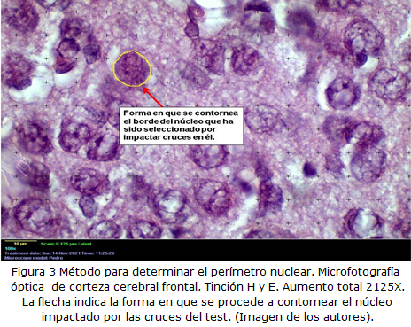
Methods: A basic experimental study of a series of cases was carried out on 20 young Wistar rats, of which 10 were descendants of pregestational diabetes. Morphometric indicators of the nervous tissue were characterized as thickness of the cortex and nuclear indicators such as perimeter.
Results: The average height of the cortical gray matter showed a value of 1.224 ± 303.7 μm for the control group and 1.014 ± 376.0 μm for the cases. When applying the mean difference test, a significant difference was found (p ≤ 0.05) in favor of the control group. The values of the measurement of the nuclear perimeter in the control group was 42.80 ± 7.23 μm and in the experimental group the average was 39.68 ± 6.52 μm. When applying the mean different test, a significant difference was found (p ≤ 0.05) at favor of control group presenting greater nuclear perimeter.
Conclusions: The greatest cortical thickness and nuclear perimeter corresponded to the control group, evidencing the deleterious effect os diabetes mellitus on neurodevelopment.
DeCS: DIABETES MELLITUS; RATS, WISTAR; GRAY MATTER; ANIMALS, LABORATORY; PREGNANCY.
Downloads
References
1. Snell RS. Estructura y localización funcional de la corteza cerebral. En: Snell RS, editor. Neuroanatomía clínica. 7ma ed. España: Wolters Kluwer Health, S.A; 2014.p.479-514. Disponible en: https://www.academia.edu/43982282/NEUROANATOMÍA_de_Snell_7ma_ed
2. Valdés Valdés A, Pérez Núñez HM, García Rodríguez RE, López Gutiérrez A. Periodo embrionario. En: Valdés Valdés A, editor. Embriología Humana. La Habana: Editorial Ciencias Médicas; 2011.p.45-54.
3. Junqueira LC, Carneiro J. Histología Básica Texto y Atlas. 12ª ed. Madrid: Editorial Médica Panamericana; 2015.
4. International Diabetes Federation. Atlas de la Diabetes de la FID. 9na ed [Internet]. Bruselas: FID; 2019 [citado 19 Ene 2021]. Disponible en: https://www.diabetesatlas.org/upload/resources/material/20200302_133352_2406-IDF-ATLAS-SPAN-BOOK.pdf
5. American Diabetes Asociation. Diabetes. Estándares para la atención médica de la diabetes 2 [Internet] 2019. [citado 09 May 2021]:[aprox. 194 p.]. Disponible en: https://www.redgdps.org/los-standards-of-medical-care-in-diabetes-2019
6. Cruz Hernández J, Hernández García P, Grandía Guzmán R, Lang Prieto J, Isla Valdés A, González Padilla K, et al. Consideraciones acerca de la diabetes mellitus durante el embarazo. Rev cuba endocrinol [Internet]. 2015 Ene-Abr [citado 09 May 2019];26(1):47-65. Disponible en: http://scielo.sld.cu/scielo.php?script=sci_arttext&pid=S1561-29532015000100005
7. Vigil-De Gracia P, Olmedo J. Diabetes gestacional: conceptos actuales. Ginecol Obstet Méx [Internet]. 2017 Jun [citado 19 Ene 2021];85(6):380-90. Disponible en: https://www.medigraphic.com/pdfs/ginobsmex/gom-2017/gom176g.pdf
8. Mayo de Andrés S. Búsqueda e identificación de nuevas causas genéticas o epigenéticas de trastornos del neurodesarrollo [tesis doctoral]. Valencia: Universidad de Valencia; 2015 [citado 19 May 2021]. Disponible en: https://roderic.uv.es/bitstream/handle/10550/47942/Tesis_Mayo%20de%20Andres.pdf?sequence=1&isAllowed=y
9. Galán-López IG, Lascarez-Martínez S, Gómez-Tello MF, Galicia-Alvarado MA. Abordaje integral en los trastornos del neurodesarrollo. Rev Hosp Jua Mex [Internet]. 2017 [citado 05 May 2019];84(1):19-25. Disponible en: https://www.medigraphic.com/pdfs/juarez/ju-2017/ju171e.pdf
10. Mitanchez D. Fetal and neonatal complications in gestational diabetes:
perinatal mortality, congenital malformations, macrosomía, shoulder dystocia, birth injures, neonatal outcomes. J Ginecol Obstet Biol Reprod [Internet]. 2010 Dic [citado 09 May 2021];39(8 Suppl 2):S189-996. Disponible en: https://doi.org/10.1016/j.diabet.2010.11.013
11. Hugues Hernandorena B, Rodríguez González JC, Rodríguez García JC. Animales de laboratorio en la endocrinología: biomodelos de la diabetes mellitus tipo 1. Rev cuba endocrinol [Internet]. 2001 [citado 09 May 2021];12(3):168-77. Disponible en: http://scielo.sld.cu/pdf/end/v12n3/end06301.pdf
12. Arias-Díaz J, Balibrea J. Modelos animales de intolerancia a la glucosa y diabetes tipo 2. Nutr Hosp [Internet]. 2007 Mar-Abr [citado 19 Ene 2021];22(2):160-8. Disponible en: https://scielo.isciii.es/scielo.php?script=sci_arttext&pid=S0212-16112007000200005
13. Gorrita Pérez Y, Núñez López N, Clapés Hernández S, Fernández Romero T. Malformaciones congénitas en la descendencia de ratas diabéticas. Medimay [Internet]. 2012 [citado 19 Ene 2021];18(2):[aprox. 12 p.]. Disponible en: https://revcmhabana.sld.cu/index.php/rcmh/article/view/568/html
14. González E. Diabetes mellitus experimental: etiología de las malformaciones congénitas en descendentes de ratas diabéticas. Rev cuba endocrinol [Internet]. 2002 Ene-Abr [citado 19 May 2021];13(1). Disponible en: http://scielo.sld.cu/scielo.php?script=sci_arttext&pid=S1561-29532002000100007
15. Spalletta G, Piras F, Gili T. Brain Morphometry. Neuromethods [Internet]. 2018 [citado 10 Dic 2021];136(3):35-49. Disponible en: https://link.springer.com/book/10.1007/978-1-4939-7647-8
16. Aguilar Cordero JM, Baena García L, Rodríguez Blanque R, Latorre García J, Mur Villar N, Sánchez López AM. Diabetes mellitus materna y su influencia en el neurodesarrollo del niño; revisión sistemática. Nutr Hosp [Internet]. 2015 [citado 05 May 2019];32(6):2484-95. Disponible en: https://scielo.isciii.es/pdf/nh/v32n6/17revision12.pdf
17. Aycheh HM, Seong JK, Shin JH, Na DL, Kang B, Seo SW, et al. Biological
Brain Age Prediction Using Cortical Thickness Data: A Large Scale Cohort Study.
Front Aging Neurosci [Internet]. 2018 [citado 11 Oct 2021];10:252. Disponible en: https://www.ncbi.nlm.nih.gov/pmc/articles/PMC6113379/
18. Fernández Viadero C, Verduga Vélez R, Crespo Santiago D. Patrones de envejecimiento cerebral. Rev Esp Geriatr Gerontol [Internet]. 2017 Jun [citado 11 Oct 2021];52(Suppl 1):7-14. Disponible en: https://www.elsevier.es/es-revista-revista-espanola-geriatria-gerontologia-124-pdf-S0211139X18300738
19. Díaz Rojas P. Introducción a la Morfometría y la Estereología [Internet]. Holguín: Universidad de Ciencias Médicas de Holguín; 2016 [citado 11 Oct 2021]. Disponible en: http://uvs.ucm.hlg.sld.cu/mod/resource/view.php?id=3459
20. Ramanoel S, Hoyau E, Kauffmann L, Renard F, Pichat C, Boudiaf N, et al. Gray Matter Volume and Cognitive Performance During Normal Aging. A Voxel Based Morphometric Study. Front Aging Neurosci [Internet]. 2018 Ago [citado 11 Oct 2021];10:35. Disponible en: https://www.ncbi.nlm.nih.gov/pmc/articles/PMC6085481/pdf/fnagi-10-00235.pdf
21. Arizamendi J, Carmona Pertuz V, Colmenares A, Gómez Hoyos D, Palomo T. Diabetes Gestacional y Complicaciones Neonatales. rev fac med [Internet]. 2012 Jul-Dic [citado 29 Abr 2020];20(2):50-60. Disponible en: http://www.scielo.org.co/scielo.php?script=sci_arttext&pid=S0121-52562012000200006
22. Jevtic P, Edens LJ, Vukovic LD, Levy DL. Sizing and shaping the nucleus: mechanisms and significance. Curr Opin Cell Biol [Internet]. 2014 Jun [citado 29 Abr 2020];28:16-27. Disponible en: https://www.ncbi.nlm.nih.gov/pmc/articles/PMC4061251/
23. Seaman L, Meixner W, Snyder J, Rajapakse I. Periodicity of nuclear morphology in human fibroblasts. Nucleus [Internet]. 2015 [citado 09 May 2021];6(5):408-16. Disponible en: https://www.ncbi.nlm.nih.gov/pmc/articles/PMC4915517/
24. Toledo Hidalgo D. Indicadores morfométricos del Carcinoma papilar de tiroides en pacientes de la provincia Holguín [tesis]. Holguín: Facultad de Ciencias Médicas Mariana Grajales Coello; 2018.
25. Yamamoto JM, Benham JL, Dewey D, Sánchez JJ, Murphy H, Murphy HR, Feig DS, et al. Neurocognitive and behavioural outcomes in offspring exposed to maternal pre-existing diabetes: a systematic review and meta-analysis. Diabetología [Internet]. 2019 Sep [citado 09 May 2021];62(9):1561-74. Disponible en: https://pubmed.ncbi.nlm.nih.gov/31278412/
Published
How to Cite
Issue
Section
License
Copyright (c) 2022 Leticia Mármol-Caballero, Pedro Augusto Díaz-Rojas, Doralny Peña-Marrero, Liúdisis Silva-Jardínez, Dunia Yailín Macareño-Ávila

This work is licensed under a Creative Commons Attribution-NonCommercial 4.0 International License.
Copyright: Camagüey Medical Archive Magazine, offers immediately after being indexed in the SciELO Project; Open access to the full text of the articles under the principle of making available and free the research to promote the exchange of global knowledge and contribute to a greater extension, publication, evaluation and extensive use of the articles that can be used without purpose As long as reference is made to the primary source.
Conflicts of interest: authors must declare in a mandatory manner the presence or not of conflicts of interest in relation to the investigation presented.
(Download Statement of potential conflicts of interest)
The Revista Archivo Médico de Camagüey is under a License Creative Commons Attribution-Noncommercial-No Derivative Works 4.0 International (CC BY 4.0).
This license allows others to distribute, to mix, to adjust and to build from its work, even for commercial purposes, as long as it is recognized the authorship of the original creation. This is the most helpful license offered. Recommended for maximum dissemination and use of licensed materials. The full license can be found at: https://creativecommons.org/licenses/


 22 julio 2025