

Tomographic evaluation of cranial basals cisterns
Abstract
Introduction: The radiological characteristics of the cisterns of the base are of interest in the diagnosis, evolution and prognosis of patients with traumatic injuries. Currently, their condition is an important predictive factor in patients with severe head trauma. For the most part they refer only to normal, compressed or absent cisterns.
Objective: To determinate the normal measurements of the principal basal cisterns in a tomagraphy and their relation with age and sex.
Methods: An analytical, cross-sectional study was carried out in a period of two months: March and April 2021, in joint work of the Neurosurgery and Imaging services of the Manuel Ascunce Domenech University Hospital in Camagüey province. The universe was made up of 101 patients over 18 years of age with skull tomographies without alterations. The study included the evaluation of simple CT images of the skull of patients with suspected cerebrovascular disease, study of late-onset epilepsy, confusional syndrome or cephalalgic syndrome. The crural, interpeduncular, ambiens and quadrigeminal cistern were evaluated. The measurements were always performed by specialists in Imaging and Neurosurgery with years of experience. The basic selection criterion was that the tomography was reported without alterations, regardless of age and sex.
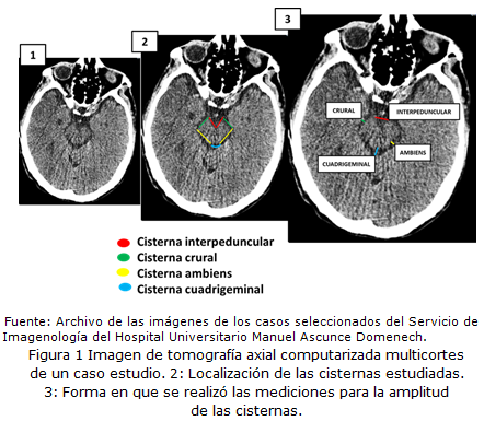
Results: The sample was 101 patients, 53 males and 48 females. The average width of the interpeduncular cistern was 5.5 mm, besides crural cistern averages 2.3 mm and the ambiens and cuadrigeminal cisterns average 3.1 mm and 5 mm respectively. The patients older than 60 years had major dimensions of the cranial cisterns.
Conclusions: The width of basal cisterns could be change with age but not with sex. This is associated with brain physiological aging.
DeCS: SUBARACHNOID SPACE; CEREBROSPINAL FLUID; BRAIN INJURIES, TRAUMATIC; INTRACRANIAL HYPERTENSION; TOMOGRAPHY, X-RAY COMPUTED.
Downloads
References
1. Díaz JF, Medina L, Herrera JM, Mural M, Baihausakas G. Cisternas Basales Cerebrales: Estudio de la Anatomía aplicado a la Resonancia magnética 3 tesla. 9na Jornadas Científicas y de Gestión [Internet]. 2016 [citado 28 Jul 2021]. Disponible en: https://repositorio.hospitalelcruce.org/xmlui/bitstream/handle/123456789/444/CISTERNAS%20BASALES%20CEREBRALES.pdf?sequence=1&isAllowed=y
2. Roldán Valadez E, Osorio Peralta S, Facha MT, Martínez López M, Taboada Barajas J. Anatomía radiológica del espacio subaracnoideo. Anales de Radiología México [Internet]. 2005 Ene-Mar [citado 28 Jul 2021];1:27-34. Disponible en: https://www.analesderadiologiamexico.com/temp/2005/1,%202005/Anrx051-06.pdf
3. Acuña M, Folgueira A. El espacio subaracnoideo y sus cisternas [Internet]. 2019 [citado 15 Ago 2021]. Disponible en: https://fmed.uba.ar/sites/default/files/2019-01/Cisternas%20subaracnoideas_0.pdf
4. López González L del R, Jerónimo Álvarez R, García Montes de Oca R, Legarreta Peña EE, Rodríguez Gutiérrez R, Gómez Hernández M. Valor pronóstico predictivo de la tomografía axial computarizada a los tres meses del trauma craneoencefálico severo [Internet]. Marianao, Cuba: Hospital Juan Manuel Márquez; 2004 [citado 15 Sep 2021]. Disponible en: https://xdoc.mx/preview/valor-predictivo-de-la-tac-en-el-tce-severo-607e542ac6958
5. Toledo JA, Namias R, Milano MJ. A Novel Automated Calculation of Basal Cistern Effacement Status on Computed Tomographic Imagine in Traumatic Brain Injury. Cureus [Internet]. 2021 Feb [citado 22 Jul 2021];13(2):e13144. Disponible en: https://www.ncbi.nlm.nih.gov/pmc/articles/PMC7937044/
6. Podolsky-Gondim GG, Cardoso R, Zucoloto Junior EL, Grisi L, Medeiros M, De Souza S, et al. Traumatic Brain Injury in the Elderly: Clinical Features, Prognostic Factors and Outcomes of 133 Consecutive Surgical Patients. Cureus [Internet]. 2021 Feb [citado 25 Dic 2021];13(2):e13587. Disponible en: https://www.ncbi.nlm.nih.gov/pmc/articles/PMC8009446/
7. Teasdale E, Cardoso E, Galbraith S, Teasdale G. CT scan in severe diffuse head injury: physiological and clinical correlations. J Neurol Neurosurg Psychiatry. 1984 Jun;47(6):600-603.
8. Gonzalo Domínguez M. Análisis anatomorradiológico de la circulación del líquido cefalorraquídeo con técnicas de imagen de última generación [tesis doctoral]. Salamanca: Universidad de Salamanca; 2015 [citado 09 Jul 2021]. Disponible en: https://dialnet.unirioja.es/servlet/tesis?codigo=78028&info=resumen
9. Maldonado M, Mendoza L, Robledo M, Giordanengo C, Bertona J, Bertona C. Estudio de Cisternas Cerebrales por Resonancia Magnética (RM) de alto campo [Internet]. Córdoba: Clínica Privada Vélez Sarsfield; 2012 [citado 22 Jul 2021]. Disponible en: http://congreso.faardit.org.ar/uploads/2012/poster/2012_137_PE_SNC.pdf
10. Sartori P, Alvarado L, Chirveches M, Urrutia M, Yampolsky B. Mediciones frecuentes en el sistema nervioso central mediante tomografía computada e imágenes de resonancia magnética. Rev argent radiol [Internet]. 2020 Ene [citado 12 Ago 2021];84(1). Disponible en: http://www.scielo.org.ar/scielo.php?script=sci_arttext&pid=S1852-99922020000100009
11. Wang DJ, Pandey SK, Lee DH, Sharma M. The Interpeduncular Angle: A Practical and Objective Marker for Detection and Diagnosis of Intracranial Hypotension on Brain MRI. AJNR Am J Neuroradiol [Internet]. 2019 Ago [citado 22 Ago 2021];40(8):1299-1303. Disponible en: https://www.ncbi.nlm.nih.gov/pmc/articles/PMC7048482/
12. Benjamini D, Iacono D, Komlosh ME, Peri DP, Brody DL, Basser PJ. Diffusse axonal injury has characteristic multidimensional MRI signature in the human brain. Brain [Internet]. 2021 Mar [citado 25 Dic 2021];144(3):800-816. Disponible en: https://www.ncbi.nlm.nih.gov/pmc/articles/PMC8041044/
13. Dulanto Deza JM. Clasificación de Marshall en la evaluación temprana de traumatismo encéfalo craneano. Hospital de Emergencia José Casimiro Ulloa 2014 [tesis]. Lima: Universidad de San Martin de Porres; 2015 [citado 22 Dic 2021]. Disponible en: https://repositorio.usmp.edu.pe/bitstream/handle/20.500.12727/1287/Dulanto_jm.pdf?sequence=1&isAllowed=y
14. Castillo de la Portilla A. Nivel de Correlación entre la Escala de Marshall y Uscanga con Escala de Glasgow en pacientes con diagnóstico de traumatismo cráneo encefálico en el Centro Médico Lic Adolfo López Mateos, en el periodo comprendido de enero del 2012 a marzo del 2012 [tesis]. Toluca: Universidad Autónoma del Estado de México; 2013 [citado 11 May 2021]. Disponible en: http://ri.uaemex.mx/bitstream/handle/20.500.11799/13889/Tesis.416957.pdf?sequence=1&isAllowed=y
15. Esquivel Miranda M, Steller Muñoz R. Análisis clínico-tomográfico de los pacientes que fallecieron por trauma craneoencefálico (TCE) en el Hospital México. Neuroeje [Internet]. 2004 [citado 15 Jul 2021];18(2). Disponible en: https://www.binasss.sa.cr/revistas/neuroeje/18n2/art1.pdf
16. Yung SK, In SA, Sung IK. Enlarged subarachnoid space on cranial ultrasound in preterm in infants: Neurodevelopmental implication. Sci Rep [Internet]. 2019 [citado 22 Jul 2021];9:19072. Disponible en: https://www.ncbi.nlm.nih.gov/pmc/articles/PMC6910979/
17. Glaister J, Shao M, Li X, Carass A, Roy S, Blitz AM, Prince JL, et al. Deformable model reconstruction of the subaracnoid space. Proc SPIE Int Soc Opt Eng [Internet]. 2018 Feb [citado 22 Jul 2021];10574:1057431. Disponible en: https://www.ncbi.nlm.nih.gov/pmc/articles/PMC6488218/
18. Wilk R, Kluczewska E, Likus W. Evaluation of subarachnoid space and subarachnoid cisterns in children and teenagers based on computed tomography studies. Pol J Radiol [Internet]. 2019 Ago [citado 15 Ago 2021];84:e295-e306. Disponible en: https://pubmed.ncbi.nlm.nih.gov/31636764/
19. Barredo Parra J, Ituarte Uriarte R, Mendiola Arza J, Escudero Martínez I, Zarranz Sarobe D, Gil Martín AR. Embolismo graso subaracnoideo: hallazgos radiológicos. Seram [Internet]. 2018 [citado 22 Jul 2021]. Disponible en: https://piper.espacio-seram.com/index.php/seram/article/view/2327
20. Douglas DB, Ro T, Toffoli T, Krawwchuk B, Muldermans J, Gullo J, et al. Neuroimaging of Traumatic Brain Injury. Med Sci [Internet]. 2019 Ene [citado 22 Jul 2021];7(1):2. Disponible en: https://www.ncbi.nlm.nih.gov/pmc/articles/PMC6358760/
21. Charry JS. Modelo de Predicción de Mortalidad y Pronóstico Neurológico en Pacientes Víctimas de Trauma Craneoencefálico en Colombia [tesis]. Colombia: Universidad de Jaén; 2017 [citado 21 Jun 2021]. Disponible en: https://tauja.ujaen.es/bitstream/10953.1/11753/1/CHARRY.CUELLAR.TFM.pdf
22. Uscanga Carmona MC, Castillo Lima JA, Arroyo Mayorga G. Hallazgos por tomografía computada en pacientes con trauma craneoencefálico, su relación con la evolución clínica y cálculo del edema cerebral. Neurol Neurocir Psiquiat [Internet]. 2005 [citado 25 Jul 2021];38(1):11-19. Disponible en: https://www.medigraphic.com/pdfs/revneuneupsi/nnp-2005/nnp051c.pdf
23. Chun KA, Manley GT, Stiver SI, Aiken AA, Phan N, Wang V, et al. Interobserver Variability in the Assessment of CT Imaging Features of Traumatic Brain Injury. J Neurotrauma [Internet]. 2010 [citado 22 Jul 2021];27(2):325-330. Disponible en: https://www.ncbi.nlm.nih.gov/pmc/articles/PMC2834438/
24. Chavero-Magro MJ, Rivera-Fernández R, Busquier-Hernández H, Fernández- Modéjar E, Pino-Sánchez F, Díaz-Contreras R, et al. Capacidad pronóstica de los signos de herniación cerebral en pacientes con afectación neurológica estructural. Med intensiva [Internet]. 2007 Ago [citado 22 Jul 2021];31(6):281-288. Disponible en: https://www.medintensiva.org/es-content-articulo-13108548
25. Cherian I, Yi G, Munakomi S. Cisternostomy: Replacien the age old decompressive hemicraniectomy. Assian J Neurosurg [Internet]. 2013 Jul-Sep [citado 22 Jul 2021];8(3):132-138. Disponible en: https://www.ncbi.nlm.nih.gov/pmc/articles/PMC3877499/
Published
How to Cite
Issue
Section
License
Copyright (c) 2022 Gretel Mosquera-Betancourt, Rogers Téllez-Isla, Elizabet Ramírez-Reyes, Luis Manuel Amador-Aguilar

This work is licensed under a Creative Commons Attribution-NonCommercial 4.0 International License.
Copyright: Camagüey Medical Archive Magazine, offers immediately after being indexed in the SciELO Project; Open access to the full text of the articles under the principle of making available and free the research to promote the exchange of global knowledge and contribute to a greater extension, publication, evaluation and extensive use of the articles that can be used without purpose As long as reference is made to the primary source.
Conflicts of interest: authors must declare in a mandatory manner the presence or not of conflicts of interest in relation to the investigation presented.
(Download Statement of potential conflicts of interest)
The Revista Archivo Médico de Camagüey is under a License Creative Commons Attribution-Noncommercial-No Derivative Works 4.0 International (CC BY 4.0).
This license allows others to distribute, to mix, to adjust and to build from its work, even for commercial purposes, as long as it is recognized the authorship of the original creation. This is the most helpful license offered. Recommended for maximum dissemination and use of licensed materials. The full license can be found at: https://creativecommons.org/licenses/


 22 julio 2025