

Giant ovarian mucinous borderline cystoadenoma, in a posmenopausic patient
Abstract
Introduction: Ovarian tumors are not as frequent as those of the uterus and breast. They constitute the third group of benign and malignant tumors in women. Border cystadenoma has clinicopathologic intermediate features between those of benign and malignant tumors.
Objective: To describe the successful therapeutic management of a case of a postmenopausal woman with borderline mucus cystadenoma of ovary.
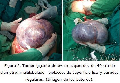
Case report: A hypertensive and diabetic patient, operated in the Lucia Iñiguez Landín Clinical Surgical Hospital in Holguin province, 54-year-old postmenopausal, with a giant tumor of the left ovary and satisfactory clinical-surgical evolution.
Conclusions: Border mucinous cystadenoma is usually limited to the ovary at the time of diagnosis, without capsular or peritoneal invasion. The risk of malignant degeneration is highly variable and is related to age. For its treatment, oophorectomy, omentectomy and biopsy of the contralateral ovary, retroperitoneal nodes, visible peritoneal nodules and cytology of peritoneal fluid are recommended.
DeCS: CYSTADENOMA, MUCINOUS/therapy; OVARY; POSTMENOPAUSE; NEOPLASMS/surgery; CASE REPORTS.
Downloads
References
1. López Carpintero N, Salazar Arquero FJ, Ibáñez Santamaría A, Fuente Valero J de la, Aramendi Sánchez T, Hernández Aguado JJ. Tumor ovárico benigno proliferante mucinoso de tipo endocervical con hiperplasia microglandular. Ginecol obstet Méx [Internet]. 2018 [citado 08 Abr 2021];86(4):281-88. Disponible en: http://www.scielo.org.mx/scielo.php?script=sci_arttext&pid=S0300-90412018000400281&lang=pt
2. Heller D, Nguyen L, Goldsmith LT. Association of cervical microglandular hyperplasia with exogenous progestin exposure. J Low Genit Tract Dis [Internet]. 2016 Abr [citado 20 Mar 2021];20(2):162-164. Disponible en: https://scholarship.libraries.rutgers.edu/discovery/fulldisplay/alma991031550137804646/01RUT_INST:ResearchRepository
3. Kurman RJ, Carcangiu ML, Herrington CS, Young RH. WHO Classification of Tumours of Female Reproductive Organs [Internet]. Lyon: International Agency for Researchon Cancer;2014 [citado 20 Mar 2021]. Disponible en: https://publications.iarc.fr/Book-And-Report-Series/Who-Classification-Of-Tumours/WHO-Classification-Of-Tumours-Of-Female-Reproductive-Organs-2014
4. Momenimovahed Z, Tiznobaik A, Taheri S, Salehiniya H. Ovarian cancer in the world: epidemiology and risk factors. International Journal of Women's Health [Internet]. 2019 Abr [citado 12 Ene 2021];11:287-299. Disponible en: https://www.researchgate.net/publication/332747210_Ovarian_cancer_in_the_world_Epidemiology_and_risk_factors
5. Taylor J, McCluggage WG. Ovarian seromucinous carcinoma: report of a series of a newly categorized and uncommon neoplasm. Am J Surg Pathol [Internet]. 2015 Jul [citado 12 Ene 2021];39(7):983-92. Disponible en: https://pubmed.ncbi.nlm.nih.gov/25723110/.
6. Forteza Sáez M, Pérez Trejo M, García Socarrás D, Almeida Arias DA. Cistoadenomamucinoso gigante de ovario de bajo grado de malignidad. Rev Cuba Obstetr Ginecol [Internet]. 2017 [citado 08 Abr 2021];43(3):119-124. Disponible en: http://revginecobstetricia.sld.cu/index.php/gin/article/view/240
7. Cortés Morera A, Ibáñez Morera M, Hernández Lara A, Garcí a Carranza MA. Cáncer de Ovario. Tamizaje y diagnóstico imagenológico. Rev Med leg Costa Rica [Internet]. Mar 2020 [citado 08 Abr 2021];37(1). Disponible en: https://www.scielo.sa.cr/scielo.php?script=sci_arttext&pid=S1409-00152020000100054
8. Javadi S,Ganeshan D,Qayyum A, Iyer R, Bhosale P. Ovarian Cancer, the Revised FIGO Staging System, and the Role of Imaging. AJR Am J Roentgenol [Internet]. 2016 Jun [citado 04 Abr 2021];206(6):1351-60. Disponible en: https://pubmed.ncbi.nlm.nih.gov/27042752/.
9. Carlson KJ, Skates SJ, Singer DE. Screening for ovarian cancer. Ann Intern Med [Internet]. 1994 Jul [citado 03 Mar 2021];121(2):124-32. Disponible en: https://pubmed.ncbi.nlm.nih.gov/8017726/.
10. Meissnitzer M, Cunha T. Update on Imaging of Ovarian Cancer. Current Radiology Reports [Internet]. 2016 [citado 04 Abr 2021];4(6):1-11. Disponible en: https://www.researchgate.net/publication/300078957_Update_on_Imaging_of_Ovarian_Cancer
11. Nyangoh Timoh K, Bendifallah S, Dion L, Ouldamer L, Leveque J. Tumores de ovario límite: Directrices de la CNGOF para la práctica clínica-valor de los marcadores tumorales. Gynecol Obstet Fertil Senol. 2020 Mar;48(3):277-86. Doi: 10.1016/j.gofs.2020.01.015.
12. Canlorbe G, Lecointre L, Chauvet P, Azais H, Fauvet R, Uzan C. Tumores ováricos limítrofes: Directrices de la CNGOF para la práctica clínica: Manejo terapéutico de las etapas iniciales. Gynecol Obstet Fertil Senol. 2020 Mar;48(3):287-303. Doi: 10.1016/j.gofs.2020.01.016.
Published
How to Cite
Issue
Section
License
Copyright (c) 2022 Orlando Martínez-Rosales, Joaquín Alejandro Solarana-Ortiz, Aniusky de los Ángeles-Ritchie

This work is licensed under a Creative Commons Attribution-NonCommercial 4.0 International License.
Copyright: Camagüey Medical Archive Magazine, offers immediately after being indexed in the SciELO Project; Open access to the full text of the articles under the principle of making available and free the research to promote the exchange of global knowledge and contribute to a greater extension, publication, evaluation and extensive use of the articles that can be used without purpose As long as reference is made to the primary source.
Conflicts of interest: authors must declare in a mandatory manner the presence or not of conflicts of interest in relation to the investigation presented.
(Download Statement of potential conflicts of interest)
The Revista Archivo Médico de Camagüey is under a License Creative Commons Attribution-Noncommercial-No Derivative Works 4.0 International (CC BY 4.0).
This license allows others to distribute, to mix, to adjust and to build from its work, even for commercial purposes, as long as it is recognized the authorship of the original creation. This is the most helpful license offered. Recommended for maximum dissemination and use of licensed materials. The full license can be found at: https://creativecommons.org/licenses/


 22 julio 2025