

Morfometric indicators in malignant skin melanoma
Abstract
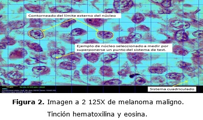
Background: malignant melanoma is an invasive tumour and difficult to treat due to its high level of mortality and aggressiveness, a morphometry study was required to provide a proper diagnosis of the disease.
Objective: to describe the behavior of morphometric indicators as nuclear area, nuclear volume and nuclear shape factor.
Methods: a case series study in 12 patients having malignant skin melanoma was carried out, in the period from September 2015 to September 2017 in Holguin Province. Theoretical and empirical methods were used; these last two methods were based on Morphometric techniques. Then a statistical analysis of the information obtained was performed and they were added on a chart.
Results: the area and nuclear volume showed low rate. The shape factor showed less levels of pleomorfism than the ones described in specialized literature.
Conclusions: when low values of nuclear volume and nuclear area in malignant melanoma are found we are dealing with hyperplastic tissue. The same happens with the incidence of the nuclear area; the values of the nuclear shape factor evidenced less pleomorfi levels than the ones described in specialized literature.
DeCS: MELANOMA/diagnosis; MELANOMA/mortality; MELANOMA/pathology; MELANOMA/therapy; SKIN NEOPLASMS/diagnosis.
Downloads
References
1. Díaz Toro YR, Gutiérrez-Ortiz R, Quimbayo Montealegre E, Jiménez-Barbosa WG. Cáncer de piel no melanoma: de la patología a la tutela. Univ Salud [Internet]. 2014 [citado 15 Mar 2018];16(2):234-244. Disponible en: http://www.scielo.org.co/pdf/reus/v16n2/v16n2a11.pdf
2. Yabor Palomo AM, Díaz Pérez M, Peña Pérez OR, Álvarez Yabor V, Morales Fontaine A. Melanoma maligno cutáneo en pacientes de la provincia de Las Tunas. Rev Electrón Zoilo [Internet]. 2015 [citado 17 May 2018];40(12):1-8. Disponible en: http://revzoilomarinello.sld.cu/index.php/zmv/article/view/483
3. Schadendorf D, van Akkooi ACJ, Berking C, Griewank KG, Gutzmer R, Hauschil A. et al. Melanoma. Lancet. 2018;392:971–84.
4. Ministerio de Salud Pública. Anuario Estadístico de Salud 2019. La Habana: Editorial Ciencias Médicas;2020.
5. Ministerio de Salud Pública. Anuario Estadístico de Salud 2017. Dirección de Registros Médicos y Estadísticas de Salud. La Habana: Ciencias Médicas;2018.
6. Hubner J, Waldmann A, Geller AC, Weinstock MA, Eisemann N, Noftz M, et al. Interval cancers after skin cancer screening: incidence, tumour characteristics and risk factors for cutaneous melanoma. Br J Cancer [Internet]. 2017 [citado 18 Mar 2019];116(2):253–259. Disponible en: https://www.nature.com/articles/bjc2016390.pdf?draft=collection
7. Ortiz Salvador JM, Ferrer DS, Saneleuterio Temporal M, Martínez V, Ferriols AP, Vilata Corell JJ, et al. Riesgo de fotocarcinogénesis asociado a la fototerapia UVB-BE. Estudio epidemiológico de un hospital terciario. Actas Dermosifiliogr [Internet]. 2018 [citado 18 May 2019];109(4):340-345. Disponible en: https://www.actasdermo.org/es-pdf-S0001731018300012
8. Iglesias-Pena N, Paradela S, Tejera Vaquerizo A, Boada A, Fonseca E. Melanoma cutáneo en el anciano: revisión de un problema creciente. Actas Dermosifiliogr [Internet]. 2019 [citado 18 May 2019];110(6):434-447. Disponible en: https://www.sciencedirect.com/science/article/abs/pii/S0001731019301231
9. Kimbrough CW, Urist MM, McMasters KM. Melanoma y neoplasias malignas cutáneas. En: Townsend CM, Beauchamp RD, Evers BM, Mattox KL, editors. Tratado de Cirugía. 20 ed. [Internet]. España: Elsevier; 2018 [citado 18 May 2019];724-753. Disponible en: https://www.clinicalkey.es/#!/content/book/3s2.0B9788491131328000305?scrollTo=%23hl0001147
10. Barros Peláez AG. Estudio Descriptivo: Diagnóstico Clínico e Histopatológico de Cáncer de Piel no Melanoma de Pacientes que Acudieron al Servicio de Dermatología del Hospital Carlo S. Andrade Marín. Rev Médica HJCA. 2015;7(2):123-127.
11. González AM, González BA, Martínez NI. Comportamiento clínico y epidemiológico del cáncer de mama en la Policlínica Alcides Pino Bermúdez. CCM [Internet]. 2012 [citado 22 May 2017];16(3):1-16. Disponible en: https://www.medigraphic.com/pdfs/correo/ccm-2012/ccm123b.pdf
12. Chuanromanee TS, Cohen JI, Ryan GL. Morphological Analysis of Size and Shape (MASS): An integrative software program for morphometric analyses of leaves. Appl Plant Scis [Internet]. 2019 [citado 22 May 2019];7(9):[aprox. 9 p.]. Disponible en: https://www.ncbi.nlm.nih.gov/pmc/articles/PMC6764432/pdf/APS3-7-e11288.pdf
13. Hevia-Montiel N, Molino-Minero-Re E, Carrillo- Bermejo AJ. Tortuosidad discreta como medida morfométrica en tumores cerebrales. Rev Mexicana Ingen Bioméd [Internet]. 2017 [citado 12 Feb 2018];38(1):189-198. Disponible en: http://www.redalyc.org/html/619/61949530014/.
14. Toledo Hidalgo D. Indicadores morfométricos del Carcinoma papilar de tiroides. Holguín. Período septiembre 2015 a septiembre 2017 [Tesis]. Holguín: Universidad de Ciencias Médicas; 2017.
15. Kashyap A, Jain M, Shukla S, Andley M. Role of nuclear morphometry in breast cancer and its correlation with cytomorphological grading of breast cancer: A study of 64 cases. J Cytol [Internet]. 2018 [citado 22 May 2018];35:41-5. Disponible en: http://www.jcytol.org/article.asp?issn=09709371;year=2018;volume=35;issue=1;spage=41;epage=45;aulast=Kashyap
16. Schirmer EC, De las Heras JI. Cancer Biology and the Nuclear Envelope: Recent Advances May Elucidate Past Paradoxe. New York: Springer; 2014.
17. Mijovic Z, Kostov M, Mihailovic D, Zivkovic N, Stojanovic M, Zdravkovic M, et al. Correlation of nuclear morphometry of primary melanoma of the skin with clinicopathological parameters and expression of tumor suppressor proteins. J BUON [Internet]. 2013;18(2):471-476.
18. Sánchez Pérez E. Caracterización histológica y morfométrica de la piel facial en personas mayores de 40 años de la provincia Holguín [Tesis]. Holguín: Universidad de Ciencias Médicas, Hospital Vladimir Ilich Lenin; 2017.
19. Azkue D, Martínez A. Variación del cariotipo, volumen nuclear y contenido de adn en siete especies de oxalis. Darwiniana [Internet]. 1984 [citado 13 Jul 2018];25(1/4):267-277. Disponible en: https://www.jstor.org/stable/23218078
20. Patología General. Respuestas celulares ante el estrés y las agresiones por tóxicos: adaptación, lesión y muerte [Internet]. España: Elsevier; 2010 [citado 13 Jul 2018]. Disponible en: https://www.berri.es/pdf/ROBBINS%20PATOLOGIA%20HUMANA%20(Con%20Acceso%20Online)/9788480869942
21. Mancera N, Smalley SMK, Margo CE. Melanoma of the eyelid and periocular skin: Histopathologic classification and molecular pathology. Surv Ophthalmol [Internet]. 2019 [citado 22 Dic 2019];64(3):272-288. Disponible en: https://www.ncbi.nlm.nih.gov/pubmed/30578807
22. Berger MF, Hodis E, Heffernan TP, Lissanu Deribe Y, Lawrence MS, Protopopov A, et al. Melanoma genomese quencingrevealsfrequent PREX2 mutations. Nature [Internet]. 2012 [citado 22 May 2015];485(7399):1-8. Disponible en: http://www.nature.com/nature/journal/v485/n7399/full/nature11071.html
23. Hernández Aragüés I, Avilés Izquierdo JA, Suárez Fernández R. Melanoma cutáneo de cabeza y cuello. Piel. Form contin dermatol [Internet]. 2019 [citado 22 May 2019];34(2):103-106. Disponible en: https://www.elsevier.es/es-revista-piel-formacion-continuada-dermatologia-21-articulo-melanoma-cutaneo-cabeza-cuello-S0213925118303320
24. García Gutiérrez M, Baldomir Mesa T, Castillo García R. Comportamiento de parámetros citológicos en el melanoma extensivo superficial. Medicentro [Internet]. 2009 [citado 03 Mar 2018];13(2):1-6. Disponible en: http://www.medicentro.sld.cu/index.php/medicentro/article/view/311
25. Cirón Martínez G, Herrera Pérez MA. Neoplasias. En: Cirón Martínez G, editor. Anatomía patológica, temas para enfermeras. La Habana: Ecimed; 2005. p. 152-199.
26. Bosserhoff AK. Melanoma Development: Molecular Biology, Genetics and Clinical Application. 2da. ed. Switzerland: Springer; 2017.
27. Sánchez I, Lloret P, Mihm M. Melanoma Maligno. En: Torres V, Camacho FM, Mihm MC, Sober A, Sánchez I, editores. Dermatología práctica ibero-latinoamericana. Atlas, enfermedades sistémicas asociadas y terapéutica. México, D.F: Galderma; 2005. p. 1359-84.
28. Song H, Tao Y, Ni N, Zhou X, Xiong J, Zeng X, et al. miR-128 targets the CC chemokine ligand 18 gene (CCL18) in cutaneous malignant melanoma progression. J Dermatol Sci [Internet]. 2018 [citado 03 Mar 2019];91(3):317-324. Disponible en: https://www.ncbi.nlm.nih.gov/pubmed/30025750
29. Schumacher M, Fukuda K. Opioids. En: Gropper MA, editors. Miller's Anesthesia. 9na ed. [Internet]. Philadelphia: Elsevier; 2020. p. 680-741. [citado 24 Feb 2020]. Disponible en: https://www.clinicalkey.es/#!/content/book/3s2.0B9780323596046000249?scrollTo=%23hl0002846
30. Joel de L, Pareja A. Inmunología del cáncer II: bases moleculares y celulares de la carcinogénesis. Horiz Med [Internet]. 2019 [citado 24 Feb 2020];19(2):84-92. Disponible en: http://www.scielo.org.pe/scielo.php?script=sci_arttext&pid=S1727-558X2019000200011&lng=es
Published
How to Cite
Issue
Section
License
Copyright (c) 2020 Yamila Oro-Pozo, Elizabeth Leyva-Sánchez, Pedro Augusto Díaz-Rojas

This work is licensed under a Creative Commons Attribution-NonCommercial 4.0 International License.
Copyright: Camagüey Medical Archive Magazine, offers immediately after being indexed in the SciELO Project; Open access to the full text of the articles under the principle of making available and free the research to promote the exchange of global knowledge and contribute to a greater extension, publication, evaluation and extensive use of the articles that can be used without purpose As long as reference is made to the primary source.
Conflicts of interest: authors must declare in a mandatory manner the presence or not of conflicts of interest in relation to the investigation presented.
(Download Statement of potential conflicts of interest)
The Revista Archivo Médico de Camagüey is under a License Creative Commons Attribution-Noncommercial-No Derivative Works 4.0 International (CC BY 4.0).
This license allows others to distribute, to mix, to adjust and to build from its work, even for commercial purposes, as long as it is recognized the authorship of the original creation. This is the most helpful license offered. Recommended for maximum dissemination and use of licensed materials. The full license can be found at: https://creativecommons.org/licenses/


 22 julio 2025