Morphometric indicators of thyroid papillary carcinoma diagnosed by escisional biopsy
Abstract
Background: thyroid is where malignant endocrine tumor diseases originate more frequently. This entity possesses multiple histological variants added to this antecedent, which are usually the cause of important diagnostic doubts. For this reason, studies in which more and more morphometric procedures are added have been developed. The success of an individualized treatment depends on an accurate diagnosis, in which morphometry is recognized by ruling out subjectivity in diagnosis, becomes a valuable tool.
Objective: morphometric characterization of papillary thyroid carcinoma diagnosed by excisional biopsies. Morphometric.
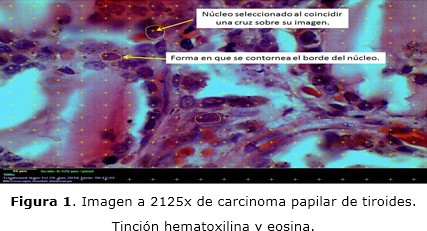
Methods: a morphometric study of a series of cases was carried out with 12 patients with this histopathological diagnosis, attended at the Vladimir Ilich Lenin University Provincial Hospital. 340 fields were selected and 10 465 cell nuclei were measured, which was our sample. Nuclear morphometric indicators were characterized, such as area, volume and shape factor.
Results: nuclear area and volume showed increased values compared to benign nodular disease values from other studies. The value of the nuclear form factor approached one, so the nuclei tend to be round and it was observed that the higher the values of the nuclear area, the higher the values of the nuclear form factor.
Conclusions: morphometric indicators of papillary thyroid carcinoma were characterized in the studied cases that may contribute to histopathological diagnosis.
DeCS: THYROID CANCER, PAPILLARY/diagnosis; THYROID CANCER, PAPILLARY/pathology; THYROID CANCER, PAPILLARY/therapy; THYROID GLAND/pathology; THYROID GLAND/anatomy&histology.
Downloads
References
1. Kumar V, Abbas AK, Aster JC. Neoplasias de la glándula tiroides. En: Robbins, editor. Patología Humana. 9na ed. España: Elsevier; 2013.
2. Ministerio de Salud Pública. Anuario Estadístico de Salud 2017. La Habana, Cuba: Dirección Nacional de los Registros Médicos y Estadístico de Salud;2019.
3. Henderson YC, Soon Hyun A, Junsun R, Yunyun C, Michelle D, El Nagar A, et al. Development and Characterization of Six New Human. J Clin Endocrinol Metab [Internet]. 2015 [citado 23 Mar 2019];100(1):1-10. Disponible en: https://watermark.silverchair.com/jcemE243.pdf
4. Sosa Martín G, Ernand Rizo S. Aspectos actuales del carcinoma bien Diferenciado de tiroides. Rev Cub Cirug [Internet]. 2016 [citado 25 Mar2019];55(1):54-66. Disponible en: http://www.medigraphic.com/pdfs/cubcir/rcc-2016/rcc161f.pdf
5. Lobo FD, Nirupama M, Pai RR, Kini AU. Cytomorphology of Warthin‑like variant of papillary thyroid carcinoma. Thyroid Res Pract [Internet]. 2015 [citado 23 Mar 2019];12(2):80-82. Disponible en: http://content.ebscohost.com/ContentServer.asp
6. Cacho Díaz B, Spínola Maroño H, Granados García M, Reyes Soto G, Cuevas Ramos D, Herrera Gómez A, et al. Metástasis cerebrales en pacientes con cáncer de tiroides. Med Int Méx [Internet]. 2017 [citado 26 Mar 2019];33(4):452-458. Disponible en:
http://www.scielo.org.mx/pdf/mim/v33n4/0186-4866-mim-33-04-00452.pdf
7. Acosta Pérez R, Hidalgo Martínez BD, Zambrano Cedeño CP, Gámez Brito D. Utilidad de los métodos diagnósticos en detección de cáncer tiroideo. Rev Ciencias Salud [Internet]. 2017 [citado 26 Mar 2019];1(2):1-10. Disponible en: http://revistas.utm.edu.ec/index.php/QhaliKay/article/view/761/604
8. Fuenzalida R, Vial I, Rojas V, Pizarro F, Puebla V, Vial G. Cirugía profiláctica en cáncer medular de tiroides hereditario. Rev Chil Cir [Internet]. 2017 [citado 26 Mar 2019];69(3):268-272. Disponible en: https://scielo.conicyt.cl/scielo.php?script=sci_arttext&pid=S0718-40262017000300017&lng=es
9. Hatice T, Ozlem E, Zeliha Esin C, Arsenal Sezgin A. Associations Between Nucleus Size, and Immunohistochemical. Pathol Oncol Res [Internet]. 2017 [citado 23 Mar 2019];25:401-408. Disponible en: https://link.springer.com/content/pdf/10.1007%2Fs12253-017-0337-9.pdf
10. Kashyap A, Jain M, Shukla S, Andley M. Role of nuclear morphometry in breast cancer and its correlation with cytomorphological grading of breast cancer: A study of 64 cases. J Cytol [Internet]. 2019 [citado 22 May 2019];35(1):41-45. Disponible en: http://www.jcytol.org/article.asp?issn=09709371;year=2019;-volume=35;issue=1;spage=41;epage=45;aulast=Kashyap
11. Sánchez Pérez E. Caracterización histológica y morfométrica de la piel facial en personas mayores de 40 años de la provincia Holguín [Tesis]. Holguín: Universidad de Ciencias Médicas, Hospital Vladimir Ilich Lenin; 2017.
12. López Pérez R, García Gutiérrez M, Pérez Pérez de Prado N, López Pérez G. Estudio histomorfométrico del núcleo celular del carcinoma papilar de Tiroides. Medicent Electrón [Internet]. 2013 [citado 18 Abr 2019];17(1):1-8. Disponible en: http://scielo.sld.cu/pdf/mdc/v17n1/mdc03113.pdf
13. Kirillov VA, Yuschenko YP, Papleuka AA, Demidchik EP. Thyroid carcinoma diagnosis based on a karyometric parameters of follicular cells. Cancer [Internet]. 2001 [citado 10 May 2019];92(7):1-10. Disponible en: http://www.ncbi.nlm.nih.gov/pubmed/11745254
14. Bhatia JK, Boruah D, Manglem R. Study of Fine Needle Aspiration Cytology of Thyroid Lesions by Morphometry. Sch J App Med Sci [Internet]. 2019 [citado 23 Mar 2019];6(7):2712-2716. Disponible en: http://www.saspublisher.com/.
15. Macedo Alessandra A, Pessoti Hugo C, Almansa Luciana F, Felipe Joaquim C, Kimura Edna T. Morphometric information to reduce the semantic gap in the characterization of microscopic images of thyroid nodules. Comp Method Programs Biomedic [Internet]. 2016 [citado 23 Mar 2019];130:162-174. Disponible en: https://www.clinicalkey.es/service/content/pdf/watermarked/1-s2.0-S016926071630236X.pdf?locale=es
16. Hend AS, Mina SN, El-Guindy Z, Omnia R, Ali D. Nuclear Morphometric Study in Different Thyroid Lesions. Int J Curr Microbiol App Sci [Internet]. 2018 [citado 18 Abr 2019];7(9):3483-94. Disponible en: https://doi.org/10.20546/ijcmas.2019.709.432
17. Lopamudra D, Shilpa G, Ruchika G, Kusum G, Kaur CG, Sompal S. Nuclear morphometry and texture analysis on cytological smears. Malaysian J Pathol [Internet]. 2017 [citado 23 Mar 2019];39(1):33-37. Disponible en: http://www.mjpath.org.my/2017/v39n1/nuclear-morphometry.pdf
18. Heidarian A, Yousefi E, Somma J. Digital Image Analysis of Nuclear Morphometry in Thyroid Fine Needle Biopsies. J American Society of Cytopathology [Internet]. 2017 [citado 18 Abr 2019];6(5):1-10 Disponible en: https://doi.org/10.1016/j.jasc.2017.06.189
19. Monappa V, Kudva R. Cytomorphologic Diversity of Papillary Thyroid Carcinoma. J Cytol [Internet]. 2017 [citado 23 Mar 2019];34(4):1-5. Disponible en: https://www.ncbi.nlm.nih.gov/pubmed/29118471
20. Huber MD, Gerace L. The size-wise nucleus: nuclear volume control in eukaryotes. J Cell Biol [Internet]. 2007 [citado 20 Abr 2019];179(4):583-4. Disponible en: https://www.ncbi.nlm.nih.gov/pmc/articles/PMC2080922/.
21. Jevtić P, Edens LJ, Vuković LD, Levy DL. Sizing and shaping the nucleus: mechanisms and significance. Curr Opin Cell Biol [Internet]. 2014 [citado 20 Abr 2019];28:16–27.
22. Mendaçolli PJ, Vilaverde Schmitt J, Amante Miot H, Brianezi G, Alencar Marques ME. Nuclear morphometry and chromatin textural characteristics of basal cell carcinoma. An Bras Dermatol [Internet]. 2015 [citado 18 Abr 2019];90(6):874-8. Disponible en: https://www.ncbi.nlm.nih.gov/pmc/articles/PMC4689077/.

Published
How to Cite
Issue
Section
License
Copyright (c) 2020 Deimarys Toledo-Hidalgo, Pedro Augusto Díaz-Rojas

This work is licensed under a Creative Commons Attribution-NonCommercial 4.0 International License.
Copyright: Camagüey Medical Archive Magazine, offers immediately after being indexed in the SciELO Project; Open access to the full text of the articles under the principle of making available and free the research to promote the exchange of global knowledge and contribute to a greater extension, publication, evaluation and extensive use of the articles that can be used without purpose As long as reference is made to the primary source.
Conflicts of interest: authors must declare in a mandatory manner the presence or not of conflicts of interest in relation to the investigation presented.
(Download Statement of potential conflicts of interest)
The Revista Archivo Médico de Camagüey is under a License Creative Commons Attribution-Noncommercial-No Derivative Works 4.0 International (CC BY 4.0).
This license allows others to distribute, to mix, to adjust and to build from its work, even for commercial purposes, as long as it is recognized the authorship of the original creation. This is the most helpful license offered. Recommended for maximum dissemination and use of licensed materials. The full license can be found at: https://creativecommons.org/licenses/


 22 julio 2025