

Primary spheno-temporal intra-osseous meningioma: a case report
Abstract
Background: ectopic meningioma, defined as those that have no connection with the dura mater, are rare. They are a rare variant and represent approximately 1 % of all intracranial meningioma.
Objective: to present a rare variant of an ectopic meningioma as a cause of proptosis and ophthalmoparesis found in a young patient.
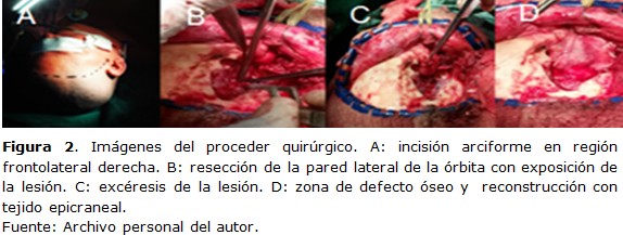
Case report: patient of 40 years of age, who 6 months ago began with pain in the right eye of moderate intensity, increased volume of the front-orbital region, decreased visual acuity and double vision. Computed tomography and MRI of the skull and orbit showed extra-axial lesion at the level of the lateral wall of the orbit with extra and intra-orbital extension with compression of adjacent structures that caused anterior displacement of the eyeball. Surgical treatment was performed with resection and the histological study concluded an ectopic meningotial meningioma of bone grade I.
Conclusions: ectopic meningioma are infrequent, surgical treatment with total resection of the lesion is the choice to avoid recurrences and may have an indication of complementary oncological treatment.
DeCS: MENINGIOMA/radiotherapy; MENINGIOMA/surgery; MENINGEAL NEOPLASMS/surgery; MARGINS OF EXCISION; EXOPHTHALMOS/surgery.
Downloads
References
1. Alcalá Cerra G, Moscote Salazar LR, Lozano Tangua CF, Sabogal Barrios R, García Quintana G. Meningioma intra diploico osteolítico primario: Reporte de caso. Rev Chil Neurocirugía [Internet]. 2010 [citado 15 Dic 2019];34(10):[aprox. 10 p.]. Disponible en:
http://www.imbiomed.com/1/1/descarga.php?archivo=Ch-Nc101-15.pdf
2. Agrawal V, Ludwig N, Agrawal A, Bulsara KR. Intra osseous intracranial meningioma. Am J Neuroradio [Internet]. 2007 Feb [citado 15 Dic 2019];28(2):[aprox. 8 p.]. Disponible en: http://www.ajnr.org/content/28/2/314
3. Ammirati M, Mirzai S, Samii M. Primary intra osseous meningiomas of the skull base. Acta Neurochir. 1990 Mar;107(1-2):56-60.
4. Daffner RH, Yakulis R, Maroon JC. Intra osseous meningioma. Skeletal Radiol. 1998 Feb;27(2):108-11.
5. Ichimura S, Hara K, Shimokawa R, Kagami H, Inaba M. A Case of Intra osseous Microcystic Meningioma Without a Mass Lesion. Neurol Med Chir (Tokyo)[Internet]. 2013 Oct [citado 16 Dic 2019];53(10):[aprox. 13 p.]. Disponible en: https://www.ncbi.nlm.nih.gov/pmc/articles/PMC4508748/.
6. Abdellaoui M, Andaloussi IB, Tahri H. Meningioma in spheno-orbital plate: report of a case with review of the literature. Pan Afr Med J [Internet]. 2015 Jun 25 [citado 16 Dic 2019];21:[aprox. 10 p.]. Disponible en:
https://www.ncbi.nlm.nih.gov/pmc/articles/PMC4546790/.
7. Mariniello G, Maiuri F, de Divitis E, Bonavolonta G, Tranfa F, Iuliano A, et al. Lateral orbitotomy for removal of sphenoid wing meningiomas invading the orbit. Neurosurgery. 2010 Jun;66(6 Suppl):287-92.
8. Amirjamshidi A, Abbasioun K, Amiri SR, Ardalan A, Hashemi SM. Lateral orbitotomy approach for removing hyperostosing en plaque sphenoid wing meningiomas. Description of surgical strategy and analysis of findings in a series of 88 patients with long-term follow up. Surg Neurol Int [Internet]. 2015 May [citado 16 Dic 2019];6:[aprox. 13 p.]. Disponible en: https://www.ncbi.nlm.nih.gov/pmc/articles/PMC4434495/.
9. Shuangshoti S, Netsky MG, Fitz-Hugh GS. Parapharyngeal meningioma with special reference to cell of origen. Ann Otol Rhinol Laryngol. 1971 Jun;80(3):464-73.
10. Simas NM, Farias JP. Sphenoid wing en plaque meningiomas: Surgical results and recurrence rates. Surg Neurol Int [Internet]. 2013 Jul [citado 16 Dic 2019];4:[aprox. 13 p.]. Disponible en: https://www.ncbi.nlm.nih.gov/pmc/articles/PMC3740617/
11. Zimny A, Sasiadek M. Contribution of perfusion-weighted magnetic resonance imaging in the differentiation of meningiomas and other extra-axial tumors: case reports and literature review. J Neurooncol [Internet]. 2011 Jul [citado 16 Dic 2019];103(3):[aprox. 11 p.]. Disponible en: https://www.ncbi.nlm.nih.gov/pmc/articles/PMC3116130/.
12. Arregui R, Ovalle R, Castillo J. Meningioma extradural de oído medio: Reporte de un caso y revisión de la literatura. Rev Otorrinolaringol Cir Cabeza Cuello [Internet]. 2017 [citado 16 Dic 2019];77(4):[aprox. 13 p.]. Disponible en:
https://scielo.conicyt.cl/pdf/orl/v77n4/0718-4816-orl-77-04-0431.pdf
Published
How to Cite
Issue
Section
License
Copyright (c) 2020 Gabriel Eduardo Alvarez-Bermudez, Rigoberto Peñones-Montero, Jorge Alejandro Casares-Delgado, Wilson Antonio Pérez-Nicolaes, Roberto del Risco-Zayas Bazán

This work is licensed under a Creative Commons Attribution-NonCommercial 4.0 International License.
Copyright: Camagüey Medical Archive Magazine, offers immediately after being indexed in the SciELO Project; Open access to the full text of the articles under the principle of making available and free the research to promote the exchange of global knowledge and contribute to a greater extension, publication, evaluation and extensive use of the articles that can be used without purpose As long as reference is made to the primary source.
Conflicts of interest: authors must declare in a mandatory manner the presence or not of conflicts of interest in relation to the investigation presented.
(Download Statement of potential conflicts of interest)
The Revista Archivo Médico de Camagüey is under a License Creative Commons Attribution-Noncommercial-No Derivative Works 4.0 International (CC BY 4.0).
This license allows others to distribute, to mix, to adjust and to build from its work, even for commercial purposes, as long as it is recognized the authorship of the original creation. This is the most helpful license offered. Recommended for maximum dissemination and use of licensed materials. The full license can be found at: https://creativecommons.org/licenses/


 22 julio 2025