

Anatomical variations in the segmental branches of the right pulmonary artery
Abstract
Background: lung cancer is considered the most lethal of all malignant tumors. It is the disease with the highest mortality for malignant neoplasias. It requires surgery in early stages; therefore the knowledge of the anatomical variations of lung arteries is of utmost importance for surgery success.
Objective: to describe the varying patterns in the branches of the right pulmonary artery taking into account origin and number.
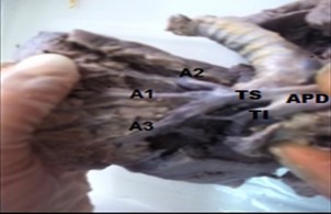
Methods: an observational and descriptive study in the Human Anatomy Department of the School of Medicine in Camagüey was conducted from November 2014 to May 2016. The universe was formed by 50 anatomical preparations of human lungs. The specimens were rinsed with pipe water and immersed in a formalin 5 % solution during a period of more than three months. A macroscopic dissection of the lungs was performed. The study variables were: number and origin of the segmental branches. A form was designed for collecting data and with the information obtained descriptive statistics and distribution of frequencies were used.
Results: the most observed variable was number. The variation of origin was less frequent. It was only found in six preparations.
Conclusions: anatomical variations of origin were less frequent, with a prevalence of non-correspondent trunk modality. Regarding number, variants in the upper lung lobe prevailed and variety corresponded to double segmental arteries, anterior segmental artery and segmental of middle lobe.
DeCS: ANATOMIC VARIATION; PULMONARY ARTERY/anatomy & histology; DISSECTION; LUNG; OBSERVATIONAL STUDY.
Downloads
References
1.García Mederos Y, Zamora Matamoros L, Sagaró del Campo N. Análisis estadístico implicativo en la identificación de factores de riesgo en pacientes con cáncer de pulmón. MEDISAN [Internet]. Ago 2015 [citado 18 Ago 2017];19(8):[aprox. 5 p.]. Disponible en: http://scieloprueba.sld.cu/scielo.php?script=sci_arttext&pid=S102930192015000800003&lng=es
2.Martínez Batista O, Moreno Infante M. Panorámica de los ensayos clínicos en el cáncer de pulmón en la provincia de Holguín. Correo cient méd holguín [Internet]. Dic 2013 [citado 18 Ago 2017];17(4):[aprox. 7 p.]. Disponible en: http://scielo.sld.cu/scielo.php?script=sci_arttext&pid=S156043812013000400001&lng=es
3.García Rodríguez ME. Estadificación y valoración mediastínica del cáncer del pulmón. Rev Cuban Cir [Internet]. Dic 2010 [citado 15 Ene 2015];49(4):[aprox. 11 p.]. Disponible en: http://scieloprueba.sld.cu/scielo.php?script=sci_arttext&pid=S003474932010000400012&lng=es
4.Marino Magdariaga DL, Marino Magdariaga E, Sagaró del Campo NM. Hallazgos anatomopatológicos en fumadoras posmenopáusicas con cánceres de mama y pulmón. MEDISAN [Internet]. Ene 2013 [citado 15 Ene 2015];17(1):[aprox. 5 p.]. Disponible en: http://scieloprueba.sld.cu/scielo.php?script=sci_arttext&pid=S102930192013000100004&lng=es
5.Díaz Toledo M, Cayón Escobar I, Crespo Díaz T, Fernández NL, Valladares CR. Quimioterapia en cáncer de pulmón avanzado en pacientes mayores de 60 años de edad del Hospital Benéfico-Jurídico (2008- 2011). Rev haban cienc med [Internet]. Abr 2014 [citado 15 Ene 2015];13(2):[aprox. 10 p.]. Disponible en: http://scieloprueba.sld.cu/scielo.php?script=sci_arttext&pid=S1729519X2014000200008&lng=es
6.Costa Montané D M, Prado Lage Y, Lozano Salazar JL, Plasencia Asorey C, Riesgo Cosme YC. Principales aspectos clínico epidemiológicos del cáncer de pulmón. MEDISAN [Internet]. Ago 2011 [citado 15 Ene 2015];15(8):[aprox. 8 p.]. Disponible en: http://scieloprueba.sld.cu/scielo.php?script=sci_arttext&pid=S102930192011000800008&lng=es
7.Hidalgo Rodríguez MT, Rojas Alonso JL, Paneque Acosta CA, Ferrer Ballagas S, Tejeda Alvares I. Presentación atípica de neoplasia de pulmón. Correo cient méd holguín [Internet]. Sep 2014 [citado 15 Ene 2015];18(3):[aprox. 7 p.]. Disponible en: http://scieloprueba.sld.cu/scielo.php?script=sci_arttext&pid=S156043812014000300019&lng=es
8.Martínez Feria F, Acosta Brooks SC, Cobián Caballero CO. Supervivencia libre de progresión de cáncer pulmonar de células no pequeñas en pacientes vacunados con CIMAvax-EGF. MEDISAN [Internet]. Dic 2015 [citado 18 Ago 2017];19(12):[aprox. 7 p.]. Disponible en: http://scieloprueba.sld.cu/scielo.php?script=sci_arttext&pid=S102930192015001200007&lng=es
9.Varona Pérez P, Torres Barbie P, Elejal de Larinaga AR, Hernández Caballero E A, NeningerVinageras E. Modelo para la prevención y manejo del cáncer de pulmón en Cuba, 2010. Rev Cuban Hig Epidemiol [Internet]. Abr 2012 [citado 15 Ene 2015];50(1):[aprox. 10 p.]. Disponible en: http://scieloprueba.sld.cu/scielo.php?script=sci_arttext&pid=S156130032012000100006&lng=es
10.Algieri R, Ottone N, Ferrante M, Bernadou M, Brofman C. Análisis del conocimiento anatómico de las estructuras del pedículo pulmonar y sus relaciones por cirujanos en formación mediante listas de chequeo.Rev Argen Anatomía Online [Internet]. Nov 2014 [citado 11 May 2016];5(4):[aprox. 7 p.]. Disponible en: http://www.anatomiaargentina.com.ar/RevArgAnatOnl2014-5%284%29p115-152-fulltext.pdf
11.Meenakshi S, Manjunath KY, Balasubramanyam V. Morphological variations of the lung fissures and lobes. Indian J Chest Dis Allied Sci [Internet]. 2004 Jul-Sep [cited 2017 May 11];[about 7 p.]. Available from: http://medind.nic.in/iae/t04/i3/iaet04i3p179.pdf
12.Şentürk A, Argüder E, Babaoğlu E, Hezer H, CananHasanoğlu H. Imagen del tromboembolismo pulmonar mediante ecografía endobronquial. Arch Bronconeumol [Internet]. Jun 2013 [citado 11 May 2017];49(6):[aprox. 3 p.]. Disponible en: http://www.archbronconeumol.org/es/diagnosticoporimagen-del-tromboembolismo/articulo/S0300289612002694/.
13.Canseco León N, Santiago Serra R.Multidetector computed angiography: a new era in the evaluation of pulmonary thromboembolism. Arch Cardiol Méx. [Internet]. 2011 Abr-Jun [cited 2017 May 11];81(2):[about 3 p.]. Available from: http://web.a.ebscohost.com/ehost/pdfviewer/pdfviewer?vid=3&sid=0c68b479fdbf-44fa-9bdf-dda8c863e1e4%40sessionmgr4010&hid=4204
14.Orts Llorca F. Anatomía humana. T III. 5ta ed. Barcelona: Editorial Científico Médica; 1980.
15.Cory R, Valentine E. Varying patterns of the lobar branches of the pulmonary artery. A study of 524 lungs and lobes seen at operation of 426 patients. Thorax [Internet]. 1959 Dic [cited 2016 May 10];[about 15 p.]. Available from: http://web.b.ebscohost.com/ehost/pdfviewer/pdfviewer?vid=3&sid=0331140772d-451e-b617-cde016253349%40sessionmgr104&hid=106
16.Warren WH, Milloy FJ. Pulmonary Vascular System and Pulmonary Hilum. Thorac Surg Clin [Internet]. 2007 [cited 2016 May 10];17:[about 16 p.]. Available from: https://www.clinicalkey.es/service/content/pdf/watermarked/1s2.0S1547412706001137.pdf?locale=es_ES
17.Sivrikoz M, Tulay C. Variations of lobar branches of pulmonary arteries in thoracic surgery patients. Surg Radiol Anat [Internet]. 2011 Ago [cited 2016 Nov 25];33(6):[about 5 p.]. Available from: http://web.ebscohost.com/ehost/pdfviewer/pdfviewer?vid=12&sid=9d4ec91bb8d0-43f3-8f6c-f094abf6a144%40sessionmgr13&hid=20
18.Ruiz Liard L. Anatomía Humana. T II. 2da ed. México: Editorial Médica Panamericana; 1989.
19.Moore KL, Dalley AF, Agur AM. Clinically Oriented Anatomy. 7th ed. USA: Lippincott Williams & Wilkins; 2014.
20.Drake RL,Wayne Vogl A, Mitchell AWW. Gray. Anatomía para estudiantes. 2da ed. Amsterdam: ELSEVIER;2010.
21.Rouviere H, Delmas A. Anatomía Humana Descriptiva, Topográfica y Funcional. TII. 10a ed. París: Editorial Masson; 2002.
22. Onuki T, Kanzaki M, Kikkawa T, Isaka T, Sakamoto K, Murasugi M, et al. New findings on the three-dimensional anatomical relations between the bronchi and pulmonary blood vessels at the pulmonary hilum. Clinical Anatomy [Internet]. 2015 May [cited 2016 May 10];28(4):[about 5 p.]. Available from: http://web.b.ebscohost.com/ehost/pdfviewer/pdfviewer?vid=3&sid=d9fdd668357f-4f7b-9c45-4f91b56b2854%40sessionmgr103&hid=125
23.Gardner E, Gray DJ, O' Rahilly R. Anatomía Humana. 3ra ed. México: Editorial Salvat; 1981.
24.Toshiteru N, Kimihiro S, Yoichi Oh, Kai O, Seiichi K, Seshiru N, et al. An analysis of variations in the bronchovascular pattern of the right upper lobe using three-dimensional CT angiography and bronchography. Gen Thorac Cardiovasc Surg [Internet]. 2015 Feb [cited 2015 May 15];63:[about 14 p.]. Available from: http://downloadv2.springer.com/static/pdf/846/art%253A10.1007%252Fs1174801505311.pdf?token2=exp=1428599154~acl=%2Fstatic%2Fpdf%2F846%2Fart%25253A1.1007%25252Fs1174801505311.pdf*~hmac=3b5bedbd8473a239533ab0a1fdbf368fd84638dfdc3845bd815424502fb8c0d
Published
How to Cite
Issue
Section
License
Copyright (c) 2018 Armando Méndez Pimentel, Sirian Saladrigas Sarduy, Iris Bacallao Cabrera, Dioneski Quesada Molina

This work is licensed under a Creative Commons Attribution-NonCommercial 4.0 International License.
Copyright: Camagüey Medical Archive Magazine, offers immediately after being indexed in the SciELO Project; Open access to the full text of the articles under the principle of making available and free the research to promote the exchange of global knowledge and contribute to a greater extension, publication, evaluation and extensive use of the articles that can be used without purpose As long as reference is made to the primary source.
Conflicts of interest: authors must declare in a mandatory manner the presence or not of conflicts of interest in relation to the investigation presented.
(Download Statement of potential conflicts of interest)
The Revista Archivo Médico de Camagüey is under a License Creative Commons Attribution-Noncommercial-No Derivative Works 4.0 International (CC BY 4.0).
This license allows others to distribute, to mix, to adjust and to build from its work, even for commercial purposes, as long as it is recognized the authorship of the original creation. This is the most helpful license offered. Recommended for maximum dissemination and use of licensed materials. The full license can be found at: https://creativecommons.org/licenses/


 22 julio 2025