

Neurofibromatosis-T1 with arteriovenous malformation and aneurysms in rapidly growing tumor mass diagnosed by angiothomograghy
Abstract
Introduction: Neurofibromatosis-type 1 is a chromosome 17 mutation induced disease. Some clinical manifestations include skin nodules, cafe au lait spots, Lisch nodules, skeletal manifestations, and cognitive deficiency. The condition can be accompanied by vascular complications; more frequent occlusion types, and less frequent hemorrhagic with arteriovenous malformation (AVM), fistulas, and aneurysms.
Objective: To present a new case of NF1 with a mass of rapid growth in the posterior cranial-cervical region with AVM, giant arterial and venous aneurysms diagnosed by multi-slice angiotomography.
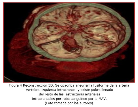
Clinical case: A female patient of 52 years of age with a history of hypertension and (NF1) comes with a tumoral mass of 15 cm, approximately 20 days of progress. The mass is situated in the posterior left cranial-cervical region that relates to mild traumatic, soft, and painful to palpation lesion, accompanied by amaurosis, cephalalgia, left facial paralysis, and transitory loss of consciousness. Multi-slice angiotomography revealed a high left vertebral arteriovenous malformation, featuring a 3 cm arteriovenous fistula with a giant venous aneurysm in the posterior occipital region. Additionally, it showed a fusiform aneurysm of the left intracranial vertebral artery and another 2 cm extracranial paravertebral aneurysm on the same side at the C1-C2 level. Surgical or interventional treatment was considered, but taking into account the patient's general condition, it was decided to follow up with symptomatic treatment.
Conclusions: In cases of rapidly growing tumor mass in patients with neurofibromatosis or plexiform neurofibroma, especially with a history of minor trauma, the possibility of vascular malformations should be excluded. Multi-slice angiotomography is a simple technique that can provide valuable information.
DeCS: NEUROFIBROMATOSIS 1; SKULL; ANEURYSM; ARTERIOVENOUS FISTULA; NEUROFIBROMA, PLEXIFORM.
Downloads
References
1. Mayo Clínic [Internet]. Rochester: Mayo Clinic; © 1998-2024 [updated 2024]; [citado 21 Jun 2024]. Neurofibromatosis. Síntomas y causas. Descripción general. Disponible en: https://www.mayoclinic.org/es/diseases-conditions/neurofibromatosis-type-1/symptoms-causes/syc-20350490
2. Duat Rodríguez A. Neurofibromatosis tipo 1. Pediatr Integral [Internet]. 2020 [citado 17 Jun 2024]; XXIV(6): 334–341. Disponible en: https://www.pediatriaintegral.es/wp-content/uploads/2020/xxiv06/05/n6-334-341_AnnaDuat.pdf
3. Mayo Clínic [Internet]. Rochester: Mayo Clinic; © 1998-2024 [updated 2024]; [citado 21 Jun 2024]. Neurofibromatosis. Diagnóstico y tratamiento. Disponible en: https://www.mayoclinic.org/es/diseases-conditions/neurofibromatosis-type-1/diagnosis-treatment/drc-20350495
4. Lavell A, Jones CW, Wong D, Counsel P, Carey-Smith R. Plexiform neurofibroma causing an ossifying subperiosteal haematoma: a rare case in the tibia of an 11-year-old girl. Skeletal Radiol [Internet]. 2017 [citado 21 Jun 2024]; 46(10): 1405-1413. Disponible en: https://pubmed.ncbi.nlm.nih.gov/28623408/https://link.springer.com/article/10.1007/s00256-017-2689-3
5. Orraca Castillo M, Licourt Otero D. Características del neurofibroma plexiforme en pacientes con neurofibromatosis tipo 1. Rev cienc méd Pinar Río [Internet]. 2012 [citado 17 Ene 2024]; 16(4): 25-35. Disponible en: http://scielo.sld.cu/pdf/rpr/v16n4/rpr04412.pdf
6. Simal-Antuña M, Fernández –Fernández C, Larrosa-Campo D. Patología neurovascular en el paciente con neurofibromatosis de tipo 1. A propósito de un caso. Rev Neurol [Internet]. 2024 [citado 17 Ene 2024]; 79(4): 115–118. Disponible en: https://pmc.ncbi.nlm.nih.gov/articles/PMC11469116/
7. Niño-Nuván, FC, Gómez-Rueda PA, Lozano-Triana CJ, Quintero EM. Neurofibroma del hipogloso en un niño colombiano. Reporte de caso. Rev Fac Med [Internet]. 2018 [citado 17 Ene 2024]; 66(1): 125-8. Disponible en: https://repositorio.unal.edu.co/bitstream/handle/unal/64976/56914-378895-3-PB.pdf?sequence=1&isAllowed=y
8. Moreno Londono MV, TaKane Imay M, González Gonzalez MC, Koga Nakamura W, Estrada Reyes CE, Islas de la Vega G. Nódulos de Lisch y ultrabiomicroscopia. Rev Mex Oftalmol [Internet]. 2014 [citado 17 Ene 2024]; 88(4). Disponible en:https://www.elsevier.es/es-revista-revista-mexicana-oftalmologia-321-articulo-nodulos-lisch-ultrabiomicroscopia-S0187451914000560
9. Muci-Mendoza R, Ramella M, Fuenmayor-Rivera D. Corkscrew retinal vessels in neurofibromatosis type 1: report of 12 cases. Br J Ophthalmol [Internet]. 2002 [citado 17 Jun 2024]; 86(3): 282-284. Disponible en: https://www.ncbi.nlm.nih.gov/pmc/articles/PMC1771041/
10. Una-Marie S, Holmes P, The London NF1 Research Group, Childs L, Roy A, Ferner RE. Neurovascular complications in adults with Neurofibromatosis type 1: A national referral center experience. Am J Med Genet [Internet]. 2022 [citado 17 Jun 2024]; 188 (10): 3009-3015. Disponible en: https://onlinelibrary.wiley.com/doi/epdf/10.1002/ajmg.a.62931
11. Brosius SN, Vossough A, Fisher MJ, Lang SS, Beslow LA, George BJ; et al. Characteristics of Moyamoya Syndrome in Pediatric Patients With Neurofibromatosis Type 1. Pediatr Neurol [Internet]. 2022 [citado 17 Jun 2024]; 134: 85-92. Disponible en:
12. Younnis M, Nasir M, Malik F, Malik SS, Malik SA. External jugular venous aneurysm: A rare case report. Radiol Case Rep [Internet]. 2024 [citado 17 Jun 2024]; 19(11): 4755-4758. Disponible en: https://pmc.ncbi.nlm.nih.gov/articles/PMC11366925/
13. Ros CA, Agorria BA, Plata JA, Canteli BS. Aneurisma sacular de vena yugular externa trombosada. A propósito de un caso. Rev Esp Cirug Oral y Maxilofac [Internet]. 2020 [citado 17 Ene 2024]; 42(4): 179-181. Disponible en: https://scielo.isciii.es/pdf/maxi/v42n4/2173-9161-maxi-42-04-00179
14. Ardhaoui H, Halily S, Mahtar M. Spontaneous evolution of a massive hematoma caused by type 1 neurofibromatosis: Case report. Int J Surg Case Rep [Internet]. 2020 [citado 17 Jun 2024]; 67: 139–141. Disponible en: https://www.ncbi.nlm.nih.gov/pmc/articles/PMC7021518/
15. Honar BN, Mirkheshti A, Memary E. Developing Cervical Hematoma Following Jaw Thrust Maneuver Triggered Diagnosis of Neurofibromatosis: A Case Report and Brief Literature Review. Anesth Pain Med [Internet]. 2017 [citado 21 Jun 2024]; 7(3): e41941. Disponible en: https://www.ncbi.nlm.nih.gov/pmc/articles/PMC5696880/
16. Correa MF, Pasik NI. Neurofibromatosis tipos 1 y 2. Rev Hosp Ital B Aires [Internet]. 2019 [citado 17 Ene 2024]; 39(4): 115-127. Disponible en: https://www1.hospitalitaliano.org.ar/multimedia/archivos/noticias_attachs/47/documentos/109202_115-127-HI5-3-19-Correa-B.pdf
17. Uchida T, Amagasaki K, Hosono A, Nakaguchi H. Neurofibromatosis type 1 with subarachnoid hemorrhage due to multiple and de novo aneurysms: a case report. J Med Case Reports [Internet]. 2021 [citado 17 Jun 2024]; 15: 411. Disponible en: https://jmedicalcasereports.biomedcentral.com/articles /10.1186/s13256-021-02967-3:
18. Vial C, Muse E, Hermida J, Lira L, Pulgar H, Várela C. Neurofibromatosis plexiforme: estudio con RM de dos casos clínicos y revisión de la literatura. Rev chil radiol [Internet]. 2007 [citado 17 Ene 2024]; 13(3). Disponible en:https://www.scielo.cl/scielo.php?script=sci_arttext&pid=S071793082007000300007
19. Macuri Valle M. Malformaciones vasculares cerebrales mediante angiotomografía cerebral en Instituto Nacional de Salud del Niño 2012-2017 [tesis]. Lima: Universidad Nacional Federico Villarreal; 2022. [citado 17 Ene 2024]. Disponible en: https://repositorio.unfv.edu.pe/bitstream/handle/20.500.13084/6015/UNFV_FTM_Macuri_Valle_Mimia_Segunda_especialidad_2022.pdf?sequence=1&isAllowed=y
20. Morita R, Abo D, Soyama T, Yoshino Y, Yoshikawa T, Kimura T; et al. Spontaneous rupture of the pancreatic arcade artery caused by neurofibromatosis type 1 successfully treated using emergency transcatheter arterial embolization, partial intra-aortic balloon occlusion, and stent graft placement: a case report and review of the literature. CVIR Endovasc [Internet]. 2020 [citado 17 Jun 2024]; 3(1): 37. Disponible en: https://pubmed.ncbi.nlm.nih.gov/32715394/
Published
How to Cite
Issue
Section
License
Copyright (c) 2025 Cristina de la Caridad Rivero-García, Cristina Fernández-Rivero, Nidhessy Pages-Morales, Brisney Mengana-Chivas

This work is licensed under a Creative Commons Attribution-NonCommercial 4.0 International License.
Copyright: Camagüey Medical Archive Magazine, offers immediately after being indexed in the SciELO Project; Open access to the full text of the articles under the principle of making available and free the research to promote the exchange of global knowledge and contribute to a greater extension, publication, evaluation and extensive use of the articles that can be used without purpose As long as reference is made to the primary source.
Conflicts of interest: authors must declare in a mandatory manner the presence or not of conflicts of interest in relation to the investigation presented.
(Download Statement of potential conflicts of interest)
The Revista Archivo Médico de Camagüey is under a License Creative Commons Attribution-Noncommercial-No Derivative Works 4.0 International (CC BY 4.0).
This license allows others to distribute, to mix, to adjust and to build from its work, even for commercial purposes, as long as it is recognized the authorship of the original creation. This is the most helpful license offered. Recommended for maximum dissemination and use of licensed materials. The full license can be found at: https://creativecommons.org/licenses/


 22 julio 2025